Visualizing Tau Turnover: STARFISH Imaging Reveals Local Protein Life Cycles
Researchers at the Max Planck Institute use the novel STARFISH method to map the simultaneous synthesis and degradation of Tau proteins within single neurons.
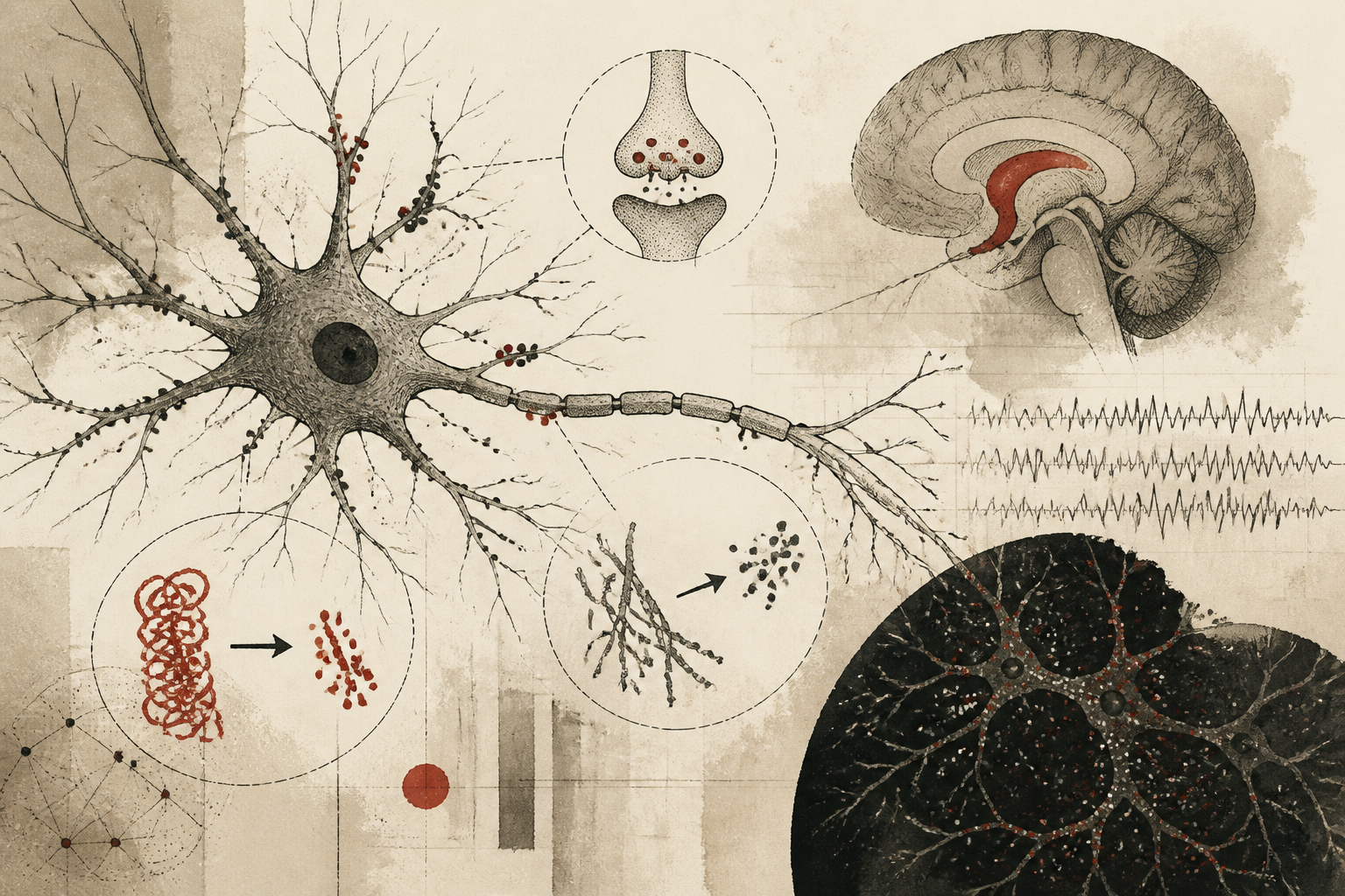
Traditional neurobiology has often viewed the neuron as a centralized factory where proteins are manufactured in the cell body and shipped to distant terminals. However, a groundbreaking study published in *Nature Neuroscience* by researchers at the Max Planck Institute for Brain Research challenges this static view by providing a high-resolution look at the life cycle of the Tau protein. Utilizing a sophisticated new imaging technique dubbed STARFISH, the research team has successfully visualized where endogenous Tau is synthesized and where it is broken down within the complex architecture of a single living neuron. This dual-tracked observation offers unprecedented insight into the delicate balance of protein homeostasis, or proteostasis, which is essential for maintaining brain health and preventing neurodegenerative decline.
Tau has long been a focal point of clinical interest due to its association with Alzheimer’s disease and other tauopathies. In a healthy brain, Tau stabilizes microtubules, which serve as the structural scaffolding and transport tracks of the cell. In diseased states, however, the protein becomes misfolded and forms toxic aggregates. Until now, tracking the 'turnover'—the continuous replacement of old proteins with new ones—of native, non-modified Tau within individual cells remained an elusive technical challenge. The development of STARFISH (Subcellular Turnover Analysis by Re-incorporation of Fluorescently-labeled In-situ Hybridization) represents a significant leap forward, allowing scientists to pinpoint the exact locations of these molecular events without relying on invasive over-expression of artificial protein variants.
The Mechanisms of Localized Proteostasis
The fundamental discovery articulated in the *Nature Neuroscience* report is that Tau is not merely a passive passenger in the distal parts of the cell. Instead, the researchers found that Tau is actively synthesized within the dendrites—the branch-like structures that receive signals from other neurons. This dendritic translation suggests that the neuron maintains a localized 'on-demand' production system to ensure that the structural integrity of the dendrite can be adjusted rapidly in response to synaptic activity. By tagging the messenger RNA (mRNA) and the resulting protein products simultaneously, the Max Planck team demonstrated that the machinery for protein synthesis is distributed far more widely than previously confirmed for this specific protein.
Crucially, the study also mapped the degradation side of the equation. Using STARFISH, the team observed that the breakdown of Tau is mediated by the neuroproteasome, a specialized protein-cleaving complex. This degradation does not occur uniformly throughout the cell; rather, it happens in specific 'hotspots' within the dendrites and the cell body. The researchers found that the rate of Tau synthesis and degradation is tightly coupled in healthy neurons. This suggests that the cell employs a sophisticated spatial sensing mechanism to ensure that the concentration of Tau remains within a narrow, functional range. When this balance is disrupted, either by excessive production or faulty waste disposal, the risk of protein aggregation increases, potentially triggering the cascade of events that leads to neurodegeneration.
Methodological Innovation: The STARFISH Framework
To achieve these results, the researchers had to overcome the limitations of traditional pulse-chase labeling, which often lacks the spatial resolution to distinguish between newly formed proteins and those that have migrated from other parts of the cell. The STARFISH technique combines metabolic labeling with high-sensitivity fluorescence microscopy. By introducing specific molecular tags that are incorporated into proteins as they are being built, and then quenching or identifying those tags during the degradation process, the team could create a color-coded map of protein age and location. This allowed for the observation of 'endogenous' Tau—meaning the protein naturally produced by the cell's own genetic instructions, rather than a foreign gene introduced by researchers.
This distinction is vital. Many previous studies relied on 'over-expression' models where the cell is forced to produce massive amounts of a fluorescently tagged protein. While useful, these models can overwhelm the cell's natural systems, leading to artifacts that do not reflect true biological behavior. By focusing on endogenous Tau, the Max Planck study provides a more accurate reflection of how the brain operates under normal conditions. The spatial maps generated by STARFISH revealed that while synthesis is widespread, the degradation of Tau via the neuroproteasome is particularly active near synaptic sites, suggesting that the life cycle of Tau is intimately linked to the neuron’s communication functions.
Limitations and Future Inquiries
Despite the power of the STARFISH imaging system, the researchers acknowledge several limitations that must be addressed in subsequent studies. First, the current research was primarily conducted in cultured neuronal models. While these models are highly controlled and allow for clear visualization, they do not fully replicate the three-dimensional complexity of a living brain with its diverse mix of glial cells and fluctuating blood flow. Future work will need to determine if the localized turnover rates observed in vitro hold true in the more chaotic environment of an intact nervous system. Furthermore, the study focused on healthy Tau; the researchers have yet to fully map how the turnover dynamics shift during the very earliest stages of tauopathy, before visible plaques or tangles appear.
Another open question concerns the specific triggers that signal the neuroproteasome to degrade a particular Tau molecule. While the study identifies the 'where' and the 'how' of degradation, the 'why'—the specific molecular flags that mark a Tau protein as 'old' or 'damaged'—remains to be fully decoded. Understanding these signals could provide new targets for therapeutic intervention. If scientists can learn how to artificially stimulate the neuroproteasome to clear out aging Tau more efficiently, they might be able to prevent the accumulation of toxic proteins that characterizes Alzheimer’s disease.
Clinical Implications for Neurodegeneration
The findings from the Max Planck Institute have significant implications for how we understand and eventually treat neurodegenerative diseases. By establishing a baseline for healthy Tau turnover, this research provides a template against which diseased states can be measured. It suggests that the loss of 'spatial control' over protein synthesis and degradation may be a precursor to disease. If Tau begins to be produced in areas where the degradation machinery is absent, or if the neuroproteasomes in the dendrites become sluggish, the resulting 'protein congestion' could lead to the formation of the deadly tangles seen in clinical cases.
Furthermore, the success of the STARFISH method opens the door to studying other proteins associated with cognitive decline, such as alpha-synuclein in Parkinson’s disease or amyloid-beta. The ability to visualize the birth and death of these proteins in real-time offers a new window into the microscopic life of the mind. As we move toward an era of personalized medicine, techniques that allow us to monitor the health of individual neurons could eventually lead to earlier diagnoses and more precise treatments that target the molecular roots of brain aging rather than just the symptoms.
Quick answers
- What is the STARFISH imaging method in neuroscience?
- STARFISH is a new technique used to visualize the life cycle of endogenous proteins within a single cell, allowing researchers to see where proteins like Tau are simultaneously created and destroyed.
- Why is Tau protein turnover important for Alzheimer's research?
- Tau helps stabilize neurons, but its accumulation into tangles is a hallmark of Alzheimer's. Understanding how the cell naturally builds and clears Tau helps identify where this process fails in disease.
- Where does Tau protein synthesis occur in a neuron?
- Contrary to older models of centralized production, new research shows that Tau is synthesized locally within the dendrites, the branches that receive signals from other neurons.
Rewritten by Zeit editorial AI. Based on original reporting at Nature Neuroscience.