Revising Neuroanatomy: The Pearl-Like Architecture of the Neuronal Axon
Recent findings challenge a century of neuroscience by revealing that axons are not smooth tubes but dynamic, beaded structures that regulate signal velocity.
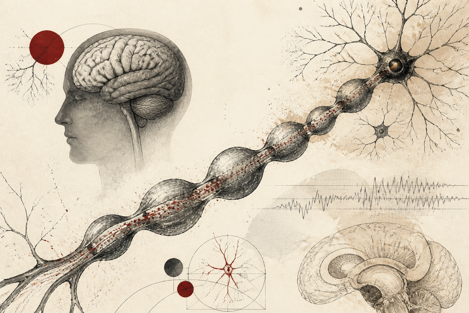
For over one hundred years, the fundamental iconography of the brain has remained largely static within the pages of academic textbooks. The standard representation of a neuron includes a cell body, branching dendrites, and a singular, long, smooth tubular projection known as the axon. This cylindrical structure has long been viewed as a biological equivalent to a copper wire—a passive conduit designed to ferry electrical impulses across the vast distances between neural circuits. However, a groundbreaking discovery reported by ScienceDaily suggests that this fundamental visualization is fundamentally flawed. Modern imaging techniques have revealed that axons do not naturally possess the smooth, uniform surface depicted in classical diagrams; instead, they resemble a series of microscopic pearls strung together. This structural revelation, which corrects a century-old misconception, implies that the physical architecture of the brain is far more fluid and functionally complex than previously understood.
The Persistence of the Tubular Paradigm
The history of neuroanatomy is built upon the work of early pioneers like Santiago Ramón y Cajal, who utilized silver staining techniques to map the intricate networks of the brain. While these early efforts provided the blueprint for modern neuroscience, the limitations of early microscopy often necessitated a degree of simplification. The assumption that axons were smooth tubes became an established truth within the field, partly because the preservation and preparation of brain tissue for imaging often caused cellular structures to collapse or distort. Consequently, the "smooth tube" became a standardized mental model for researchers and students alike. This new research indicates that the pearl-like deformations, previously dismissed by some as artifacts of tissue preparation or signs of pathology, are actually the natural, healthy state of the axon in vivo. The discovery highlights the risks of scientific inertia, where a century-old illustration can limit the scope of investigation into how nerve fibers actually behave.
Quantifying the Pearl-Like Morphology
According to the original report from ScienceDaily, scientists have observed that these bulbous, pearl-like structures are ubiquitous along the length of the axon. Rather than a steady diameter, the axon undergoes periodic expansions and contractions. This morphological variation is not static. One of the most significant findings of the study is that these "pearls" are dynamic; they can alter their shape and volume in direct response to neural activity. This suggests that the axon is not merely a structural cable but a living, responding component of the neuron. The ability of the axon to change its physical dimensions in real-time provides a new layer of complexity to our understanding of neuroplasticity. While traditional plasticity focuses on the strengthening or weakening of synapses, this discovery introduces the concept of structural axonal plasticity as a factor in brain function.
Mechanisms of Signal Velocity Regulation
The functional implications of this discovery are profound, particularly concerning the speed of information processing. In the traditional model, the velocity of an action potential—the electrical signal that travels down the axon—was thought to be determined primarily by the diameter of the tube and the presence of myelin insulation. By introducing a pearl-like geometry, the researchers have identified a new mechanism for controlling signal timing. The expansions and contractions of these structures appear to influence the rate at which electrical impulses transit the fiber. Because these shapes can change based on activity, the brain may possess the ability to self-regulate the timing of its own communications by physically reshaping its axons. This fine-tuning of signal arrival is critical for tasks requiring high-precision timing, such as sensory integration, motor coordination, and complex cognitive processing.
Methodological Breakthroughs and Limitations
This shift in perspective was made possible by advances in high-resolution imaging and live-cell observation techniques that allow researchers to see the brain in a more natural state. By avoiding the harsh fixation processes that historically smoothed out or damaged the delicate axonal membranes, the team was able to witness the beaded structure in its active environment. However, as with any transformative discovery, there are significant limitations and open questions that the scientific community must address. It remains unclear exactly what molecular mechanisms drive the formation and deformation of these pearls. Furthermore, while the study indicates that these structures help control signal speed, the precise mathematical relationship between pearl volume and conduction velocity requires further rigorous testing. Researchers also need to determine if these structures are uniform across all types of neurons or if specialized cells in different brain regions exhibit distinct axonal morphologies.
The Future of Neuroanatomical Research
The realization that a basic brain structure has been misdrawn for a century serves as a powerful reminder of the evolving nature of science. For educators and researchers, this finding necessitates a complete revision of pedagogical materials. Beyond the classroom, the implications for clinical medicine are substantial. If the pearl-like structure of the axon is essential for healthy signal conduction, then deviations from this morphology may play a role in neurodegenerative diseases or developmental disorders. Conditions characterized by "slowed" cognitive processing or communication breakdowns between brain regions may eventually be linked to the malformation or rigidity of these axonal pearls. By redefining the axon as a dynamic, beaded regulator of electricity rather than a static pipe, this research opens a new frontier in the study of how the human mind coordinates its nearly infinite complexity. As we move forward, the focus will shift from mapping the connections of the brain to understanding how the very shape of those connections dictates the speed of thought itself.
Quick answers
- How does the new discovery change our view of brain axons?
- It reveals that axons are not smooth, static tubes but dynamic, pearl-like structures that can change shape to regulate the speed of electrical signals.
- Why were axons previously thought to be smooth tubes?
- A century of textbook illustrations and limitations in early tissue preservation techniques led researchers to believe the cylindrical shape was the natural state of the axon.
- What is the functional purpose of the pearl-like shapes on axons?
- These structures appear to act as a mechanism for controlling how quickly electrical impulses travel through the brain, allowing for the fine-tuning of neural communication.
Rewritten by Zeit editorial AI. Based on original reporting at ScienceDaily — Mind & Brain.