Quiet Innovation: The Evolution of Silent Magnetic Resonance Imaging
Recent breakthroughs in acoustic reduction for neuroimaging promise to transform the patient experience and clinical accuracy in cognitive research.
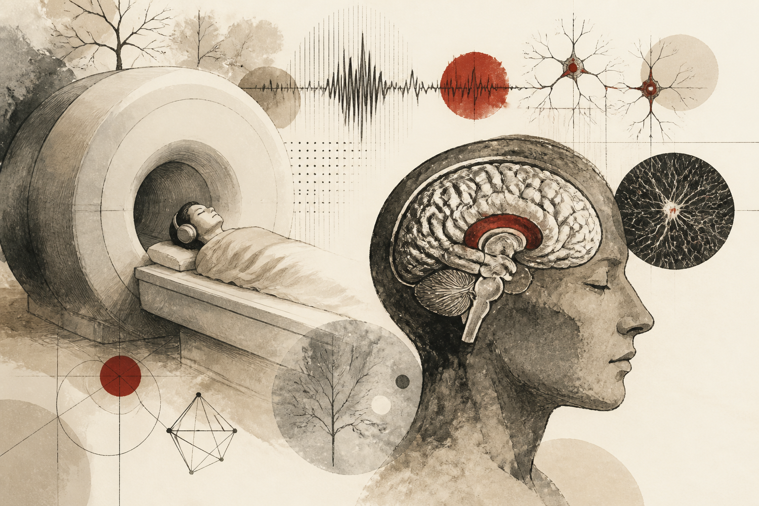
The traditional environment of neuroimaging has long been characterized by a distinctive, often distressing, auditory signature. For decades, patients and research subjects undergoing Magnetic Resonance Imaging (MRI) have had to contend with rhythmic, high-decibel knocking and buzzing sounds that can exceed 110 decibels—equivalent to the noise level of a live rock concert or a jet engine at close range. These acoustic disturbances are not merely a matter of patient discomfort; they represent a significant methodological hurdle in the fields of cognitive neuroscience and clinical diagnostics. However, a recent analysis published in Nature Neuroscience, titled "A silent leap in neuroimaging," suggests that the field is entering a transformative era where the 'silent scan' is no longer a theoretical ideal but an impending clinical reality.
The Mechanical Origin of Acoustic Disturbance
To understand the magnitude of this technological shift, one must first examine why MRI machines are traditionally loud. The noise is a byproduct of the physical laws governing electromagnetic induction. Inside the scanner, gradient coils are subjected to rapid pulses of electrical current within a strong static magnetic field. This interaction generates Lorentz forces, which cause the coils to vibrate against their mountings. Because these pulses occur thousands of times per second, the vibrations manifest as loud, rhythmic acoustic noise. Historically, this noise was accepted as an unavoidable physical consequence of high-resolution imaging. Researchers and clinicians have traditionally relied on passive protection, such as earplugs and specialized headphones, to mitigate the impact on the subject. Yet, these measures do not eliminate the bone-conducted vibrations that patients feel, nor do they address the physiological stress response triggered by such an intense auditory environment.
Foundational Findings in Acoustic Mitigation
The report in Nature Neuroscience highlights how new engineering paradigms are successfully decoupling the imaging process from the noise generation process. The core findings center on two primary innovations: hardware redesign and sophisticated pulse sequence programming. By utilizing "silent" gradient coils—which are vacuum-sealed or designed with specialized geometries to minimize vibration—and implementing "Lo-Hi" frequency sweeps in the imaging sequences, engineers have managed to reduce noise levels to just above the ambient sound of a quiet room. These advancements represent a significant leap forward because they achieve silence without sacrificing the spatial or temporal resolution required for high-stakes neurological research. The study emphasizes that these "silent" sequences allow for the monitoring of brain activity in a state that more closely mirrors natural rest or specific task-based engagement, free from the confounding variables introduced by external auditory stress.
Methodological Implications for Cognitive Research
The mechanism behind these silent leaps involves a fundamental shift in how data is sampled in the k-space—the mathematical domain where MRI raw data is stored. Traditional scans utilize rapid, jerky movements through k-space, which necessitates sudden changes in current and thus creates loud noise. The newer, quieter methods utilize a continuous, radial, or spiral trajectory. This allows the gradient coils to operate with much smoother transitions, effectively eliminating the percussive sound. For cognitive psychologists and neuroscientists, this methodology is revolutionary. In studies of auditory processing, for instance, the noise of the scanner often masks the stimuli being presented to the subject. By removing the background noise at the source, researchers can now present subtle auditory cues—such as whispers or complex linguistic tones—without interference, leading to a much higher signal-to-noise ratio in functional data.
Interpretation and Patient Impact
The interpretation of these findings suggests a move toward more ethical and inclusive neuroimaging. The acoustic environment of a standard MRI is often a barrier for specific populations, including pediatric patients, individuals with autism spectrum disorder who may have sensory sensitivities, and elderly patients with dementia who may become disoriented by loud, unfamiliar noises. The shift toward silent imaging protocols democratizes access to high-quality diagnostics. Furthermore, from a neurophysiological perspective, the absence of noise reduces the activation of the amygdala and other stress-related regions of the brain. This means that functional MRI (fMRI) data gathered in a silent environment is a more accurate representation of a subject’s baseline neural state, as it is not contaminated by the brain’s involuntary reaction to a threatening or uncomfortable environment.
Limitations and Open Questions
Despite the optimism surrounding these developments, the transition to silent imaging is not without its limitations. One primary concern involves the trade-off in acquisition time. In some current silent protocols, the time required to gather a full set of images can be longer than traditional “loud” sequences. This presents a challenge in clinical settings where throughput is a priority. Additionally, while the acoustic noise is reduced, the heat generated by the coils remains a factor that must be managed. There are also open questions regarding the scalability of these technologies. High-field magnets, such as 7-Tesla scanners used in advanced research, present even greater acoustic challenges that current silent technology is still working to overcome. Future research must determine if these silent methods can be standardized across different scanner manufacturers to ensure that data remains comparable across multi-site longitudinal studies.
The Future of Neuroimaging Excellence
The broader significance of this "silent leap" cannot be overstated. As the Nature Neuroscience report indicates, the ability to peer into the human brain without the interference of a harsh auditory environment marks a new frontier for the discipline. For institutions like Zeit Psychology Online University, these developments underscore the importance of integrating engineering breakthroughs with psychological research. As we move toward a future where the MRI suite is as quiet as a library, the quality of our data will improve, the comfort of our patients will increase, and our understanding of the human mind will become clearer. This technological evolution ensures that the next generation of neuroimaging will be defined not by the volume of the machine, but by the precision and humanity of the science it enables.
Quick answers
- Why are traditional MRI machines so loud?
- MRI noise is caused by Lorentz forces, where electrical pulses in the gradient coils react with a strong magnetic field, causing physical vibrations that manifest as loud sound.
- How does silent MRI technology work?
- It uses specialized gradient coil designs and smoother 'k-space' sampling trajectories that avoid the sudden electrical transitions responsible for acoustic noise.
- What are the benefits of silent neuroimaging for research?
- It reduces patient stress, allows for better auditory stimulus testing, and prevents stress-related brain activity from confounding functional imaging data.
Rewritten by Zeit editorial AI. Based on original reporting at Nature Neuroscience.