Novel 3D Brain Tissue Model Enhances Research into Microglial Dynamics
Researchers have developed a reproducible three-dimensional human brain model to better study how microglia influence both healthy neurological function and disease progression.
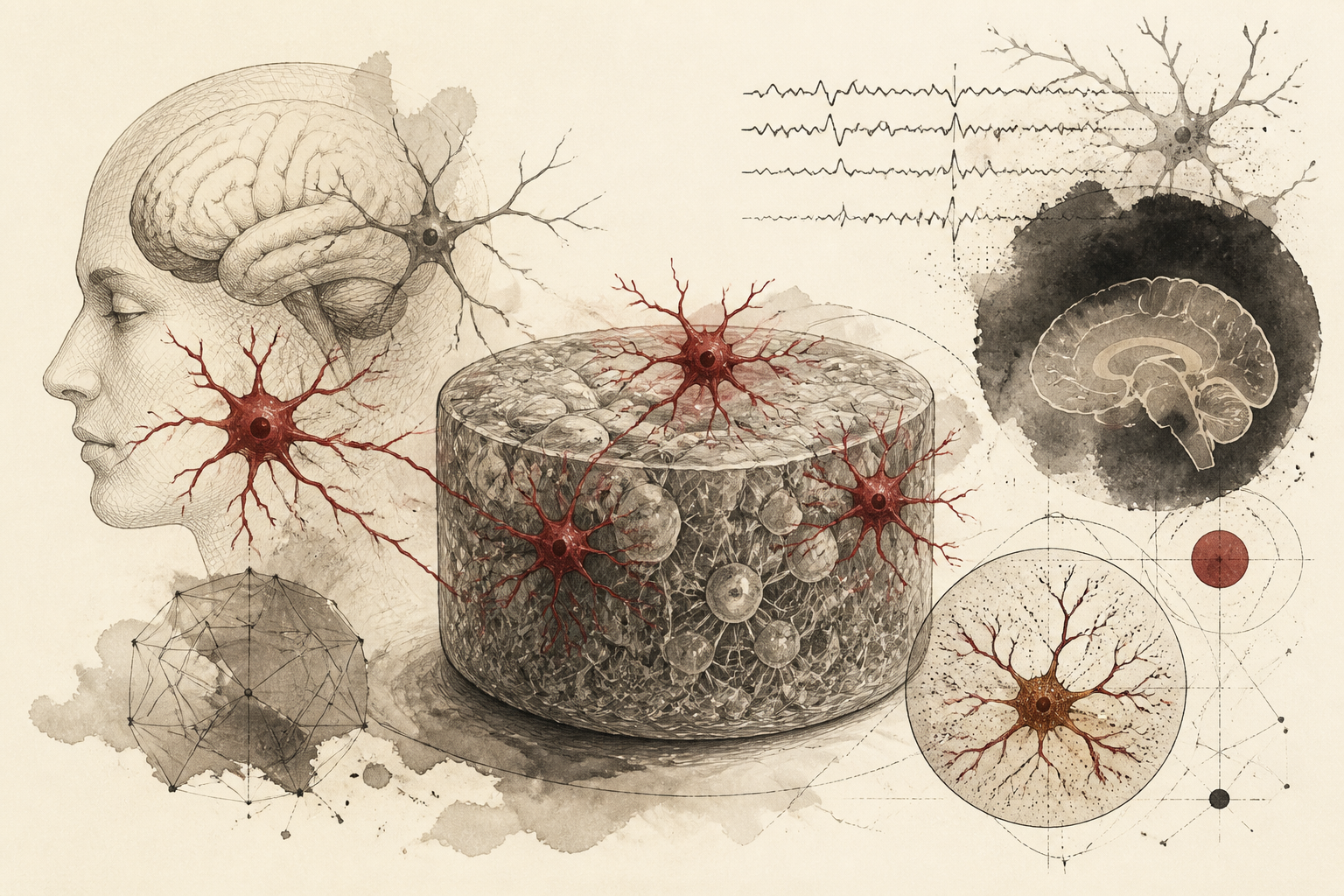
A groundbreaking advancement in neurobiology has emerged from a recent study, introducing a sophisticated three-dimensional (3D) model of human brain tissue. This innovative model is poised to revolutionize the study of microglia, the brain's primary immune cells, by providing an unprecedented view into their complex behaviors within both healthy and pathological neurological environments. Published in *Nature Neuroscience*, this research addresses a critical limitation in current neuroscience — the challenge of accurately observing microglial dynamics in a controlled yet biologically realistic setting, moving beyond the inherent limitations of traditional two-dimensional cultures. The development of this reproducible 3D framework promises to deepen our understanding of microglial contributions to neurodegenerative diseases and accelerate the identification of novel therapeutic strategies.
The Enigma of Microglia in Brain Health and Disease
Microglia are indispensable for maintaining neurological homeostasis. They continuously survey the brain microenvironment, pruning synaptic connections, clearing cellular debris, and responding to pathogens and injuries. However, these versatile cells are also central players in neuroinflammation, a process intricately linked to the initiation and progression of numerous neurodegenerative conditions. The transition of microglia from a homeostatic, surveillance state to various disease-associated phenotypes is a critical area of investigation. Understanding the factors that drive these phenotypic shifts is paramount for developing effective interventions.
Historically, studying microglia has been constrained by methodological challenges. *In vivo* studies, while offering biological realism, present difficulties in precise control and real-time observation of individual cellular behaviors. Conversely, traditional *in vitro* two-dimensional cell cultures, while providing controlled environments, often fail to recapitulate the intricate structural complexity and cellular interactions characteristic of the human brain. This fundamental disconnect frequently leads to discrepancies between laboratory observations and the complex realities observed in clinical settings, hindering the translation of research findings into clinical applications. The absence of a physiologically relevant 3D context can alter cell morphology, gene expression, and functional responses, thereby limiting the predictive power of such models.
A Reproducible 3D Model for Microglial Observation
The newly developed 3D human brain tissue model represents a significant methodological leap. This bioengineered platform is specifically designed to mimic the intricate spatial architecture of human neural tissue, thereby creating a microenvironment that more closely resembles the *in vivo* brain. By providing this authentic microenvironment, the model enables a more precise examination of microglial phenotypes – the observable characteristics that arise from the complex interplay of their genetic makeup and environmental influences. The capacity for detailed observation within this realistic setting allows researchers to track how these dynamic immune cells respond to various stimuli, ranging from subtle changes in the local environment to the overt presence of disease markers.
A key advantage of this 3D model is its high degree of reproducibility. In scientific research, reproducibility is foundational for validating findings, ensuring the reliability of experimental outcomes, and facilitating the comparison of results across different studies. This consistent performance is particularly crucial when evaluating potential therapeutic compounds or investigating the delicate mechanisms underlying neuroinflammation. The ability to consistently generate identical biological contexts allows for robust drug screening and a more confident interpretation of microglial responses to pharmacological interventions or genetic manipulations.
Unveiling Microglial Mechanisms and Translational Potential
The research demonstrates that this 3D platform allows scientists to observe not only how microglia respond to stimuli but also how they transform in the presence of disease markers. This capability is critical for unraveling the complex signaling pathways and cellular interactions that drive microglial activation and dysfunction in neurological disorders. By offering a more accurate representation of the cellular milieu, the model facilitates a deeper understanding of the processes by which microglia transition from their beneficial roles to states that contribute to neuronal damage and disease progression.
This advancement serves as a vital bridge between simplified cellular studies and the complexities of human clinical trials. Historically, the gap between preclinical findings and successful clinical translation has been wide, often due to the limitations of *in vitro* and animal models in fully capturing human pathophysiology. The human-derived nature of this 3D model, combined with its structural fidelity, significantly reduces this translational barrier. The researchers emphasize that the capacity to monitor these critical cellular shifts in a human-derived model holds substantial promise for accelerating the discovery of therapeutic targets. This includes conditions such as Alzheimer’s disease, characterized by amyloid plaque accumulation and tauopathy, and multiple sclerosis, an autoimmune demyelinating disease, where microglial activity is a prominent feature.
Limitations, Open Questions, and Future Directions
While representing a significant breakthrough, this 3D brain tissue model, like any scientific tool, possesses inherent limitations and opens new avenues for inquiry. The precise cellular heterogeneity and long-term maturation processes observed *in vivo* remain challenging to fully replicate *in vitro*, even in 3D constructs. The complexity of vascularization, crucial for nutrient and oxygen supply, and the full spectrum of intercellular communication with other brain cell types, such as astrocytes and oligodendrocytes, may require further refinement. Additionally, while the model demonstrates reproducibility, scaling up for high-throughput screening while maintaining biological fidelity could present technical challenges.
Future research will undoubtedly focus on enhancing the model's complexity to incorporate additional brain regions, diverse cell populations, and even more refined physiological cues. Exploring the integration of patient-specific induced pluripotent stem cells (iPSCs) to generate personalized disease models could further boost its translational relevance. Investigating the long-term stability and functionality of the microglial populations within these 3D constructs will also be critical for chronic disease modeling.
Implications for Psychology, Neuroscience, and Clinical Practice
For students and clinicians in psychology and neuroscience, this methodological breakthrough carries profound implications. It provides a more robust and ethically appealing alternative to animal models for studying human brain diseases, offering a direct window into human-specific cellular mechanisms. Students will benefit from this model's ability to facilitate hands-on research into complex neurobiological processes that were previously difficult to observe. Clinicians, particularly those involved in neurology and psychiatry, stand to gain from accelerated drug discovery and a deeper understanding of disease pathogenesis. The ability to test drug efficacy and predict patient responses with greater accuracy could lead to more targeted and personalized treatment approaches.
This development marks a significant step forward in the field of translational neuroscience, providing a robust tool for future investigation into the cellular basis of brain health and dysfunction. The increased confidence in preclinical findings facilitated by this model could bridge the gap between bench and bedside more effectively, bringing closer the prospect of truly effective therapies for debilitating neurological conditions. As research continues to refine and expand the capabilities of such bioengineered models, the insights gained will undoubtedly reshape our understanding of the brain and our approach to treating its disorders.
Quick answers
- What is the primary benefit of the new 3D brain model?
- The model provides a highly reproducible, three-dimensional environment that more accurately mimics human brain architecture compared to traditional 2D cultures.
- Why are microglia important in this study?
- Microglia are the brain's immune cells; understanding how they change from healthy to disease-associated states is crucial for treating neurodegenerative disorders.
- What diseases could this research impact?
- The study aims to improve understanding of conditions involving neuroinflammation and cellular dysfunction, such as Alzheimer's disease and multiple sclerosis.
Rewritten by Zeit editorial AI. Based on original reporting at Nature Neuroscience.