Mapping the Periventricular Gradient: New Insights into Multiple Sclerosis Pathology
Recent findings in Nature Neuroscience reveal how ependymal cell reactivity and adhesion loss drive the characteristic outward progression of brain lesions in Multiple Sclerosis.
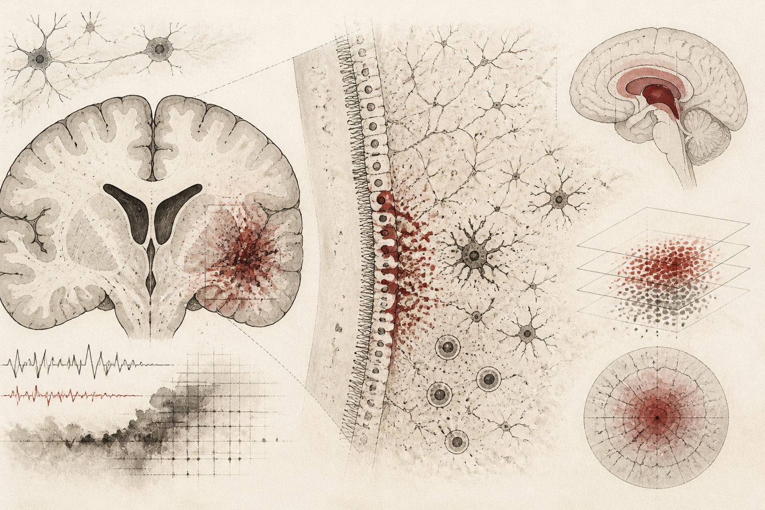
The spatial distribution of brain lesions in Multiple Sclerosis (MS) has long presented a topological puzzle for neurologists and neuropathologists. A hallmark of the disease is the concentration of tissue damage in the periventricular regions—those areas immediately surrounding the fluid-filled ventricles of the brain. While clinical observation has verified this pattern for decades, the underlying molecular drivers remained largely elusive. A landmark study published in *Nature Neuroscience*, titled "Periventricular gradient pathology is linked to ependymal reactivity and cell adhesion disruption in multiple sclerosis," provides a comprehensive map of this phenomenon. The researchers describe a distinct biological gradient where the severity of pathology decreases as the distance from the ventricular surface increases, suggesting that the cerebrospinal fluid (CSF) and the ventricular lining play a more active role in disease progression than previously understood.
The Architecture of Ependymal Disruption
At the heart of this study is the ependyma, a thin layer of ciliated epithelial cells that lines the ventricular system and serves as a critical barrier between the brain parenchyma and the CSF. The research team utilized advanced neuroimaging and high-resolution molecular profiling to examine how this barrier fails in the context of MS. They identified that the periventricular gradient is not merely a byproduct of proximity to inflammatory factors in the CSF, but rather a result of significant structural and functional changes within the ependymal cells themselves. In patients with MS, these cells exhibit what the researchers term "reactivity"—a state of cellular stress and transformation that mirrors the gliosis seen in astrocytes. This reactivity is closely linked to the breakdown of cell-to-cell adhesion molecules, which normally maintain the integrity of the ventricular lining. When these adhesive bonds are disrupted, the barrier becomes porous, potentially allowing neurotoxic substances to penetrate deeper into the brain tissue.
Investigating the Molecular Mechanism of Injury
To understand the mechanism driving this gradient, the study employed a multi-omic approach, combining single-nucleus RNA sequencing with spatial transcriptomics. By analyzing tissue samples from both MS patients and control groups, the researchers were able to pinpoint specific gene expression changes that occur at the interface of the ventricle and the white matter. The data revealed a significant downregulation of genes involved in maintaining the mechanical stability of the ependyma. Specifically, proteins that form desmosomes and tight junctions—the "glue" holding the cells together—were found to be depleted. This molecular decay precedes the full development of demyelinating lesions, suggesting that ependymal instability may be an early event in the cascade of MS pathology. The study further demonstrated that this disruption is not uniform; it follows a strict spatial gradient, with the highest levels of molecular dysregulation occurring within the first few millimeters of the ventricular wall.
Rethinking the Role of Cerebrospinal Fluid
The findings provide strong evidence for the "outside-in" hypothesis of MS progression. This theory suggests that inflammatory mediators circulating in the CSF penetrate the brain surface and initiate damage. The *Nature Neuroscience* study adds a critical layer to this theory by showing that the ependyma is not a passive casualty but an active participant. As the ependymal cells become reactive and lose their adhesive properties, they release pro-inflammatory signals that further recruit immune cells and activate microglia in the adjacent white matter. This creates a feedback loop that sustains the periventricular gradient. The researchers noted that the intensity of this gradient correlates with the overall clinical severity of the disease, suggesting that the degree of ependymal failure could serve as a biological marker for disease progression. By quantifying the spatial decline of tissue integrity, the team was able to model how the toxic environment of the CSF interacts with the vulnerable ventricular wall to shape the topography of MS lesions.
Limitations and Open Questions in Spatial Pathology
While the study offers a robust framework for understanding periventricular damage, it also highlights several areas requiring further investigation. One primary limitation is the cross-sectional nature of human post-mortem tissue analysis, which provides a snapshot of end-stage disease rather than a real-time view of the gradient's evolution. It remains unclear whether the ependymal reactivity is a primary trigger or a secondary response to pre-existing subpial or deep white matter inflammation. Additionally, while the study identifies cell adhesion disruption as a key feature, the specific triggers that initiate this molecular breakdown—be they viral, autoimmune, or metabolic—remain to be fully characterized. Future research must determine if stabilizing the ependymal barrier through pharmacological means could slow the inward migration of the disease or if the damage to the ventricular lining is irreversible once the reactive state is reached.
Implications for Future Therapeutic Strategies
The identification of a periventricular gradient linked to ependymal reactivity has profound implications for how we approach the treatment of MS. Traditional therapies have largely focused on systemic immune modulation to prevent the entry of T and B cells into the central nervous system. However, the findings from the *Nature Neuroscience* study suggest that targeting the ventricular interface could be a complementary strategy. If the ependyma can be shielded from CSF-borne inflammatory factors, or if its adhesive integrity can be reinforced, it may be possible to contain the spread of lesions within the periventricular space. This research shifts the focus toward the "borders" of the brain, highlighting the ventricular lining as a critical frontier in neuroimmunology. As we move toward more personalized medicine, understanding the specific molecular signatures of the periventricular gradient may allow clinicians to better predict disease trajectories and develop interventions that preserve the structural integrity of the brain’s internal barriers.
Quick answers
- What is the periventricular gradient in Multiple Sclerosis?
- It is a pattern where brain tissue damage is most severe near the fluid-filled ventricles and decreases in intensity as the distance from these ventricles increases.
- How do ependymal cells contribute to MS progression according to the study?
- Ependymal cells, which line the ventricles, become reactive and lose their adhesive properties, weakening the barrier and allowing inflammatory factors to damage surrounding brain tissue.
- What new technology was used to map these brain changes?
- The researchers utilized single-nucleus RNA sequencing and spatial transcriptomics to identify specific molecular changes at the interface of the brain and cerebrospinal fluid.
Rewritten by Zeit editorial AI. Based on original reporting at Nature Neuroscience.