Mapping the Neural Circuitry of Maternal Vigilance: How the Brain Wakes to Infant Cries
Recent research in Nature Neuroscience reveals a specific corticohypothalamic pathway that allows the maternal brain to selectively prioritize infant distress signals during sleep.
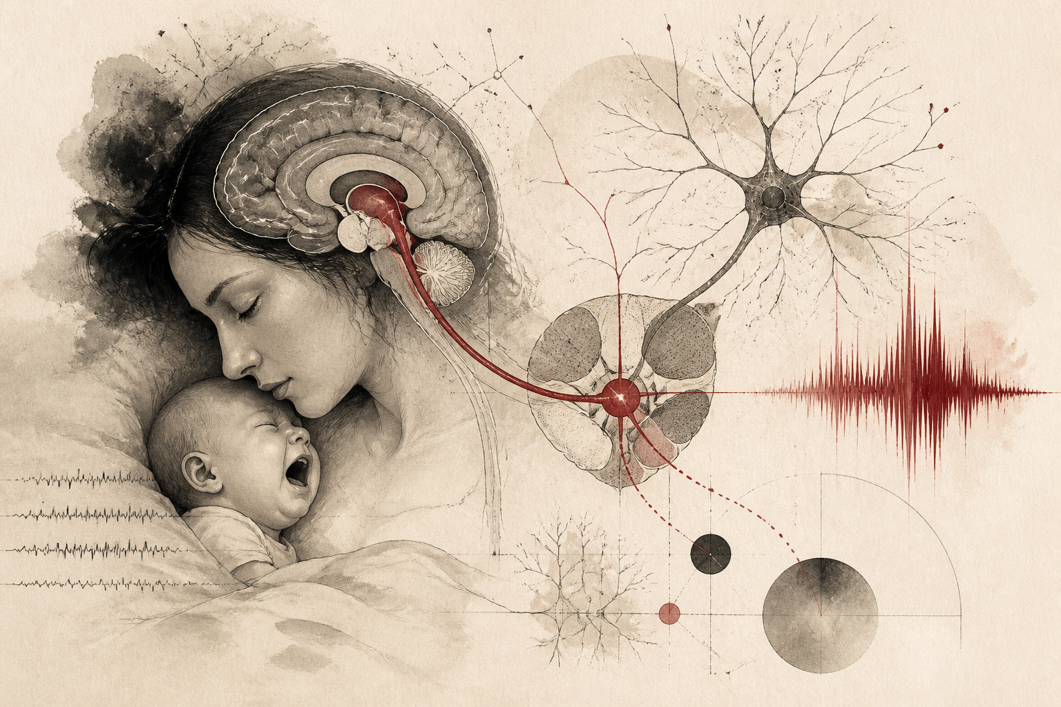
In the complex landscape of mammalian survival, few behaviors are as critical or as biologically ingrained as the maternal response to infant distress. While sleep is a state typically characterized by reduced responsiveness to external stimuli, a biological paradox exists in which mothers often remain hypersensitive to the specific sounds of their offspring. This selective arousal allows for a rapid transition from deep rest to active caregiving, yet the precise neurological architecture facilitating this filter has long remained elusive. A comprehensive study recently published in Nature Neuroscience, titled "A corticohypothalamic circuit that mediates maternal awakening to pup cries," offers a definitive map of the neural pathways responsible for this phenomenon. The researchers have identified a specialized circuit connecting the cortex to the hypothalamus, providing a mechanical explanation for how the brain maintains a high-priority monitoring system even during periods of unconsciousness.
The Architecture of Selective Arousal
The ability to ignore ambient noise while waking instantly to a specific, significant sound is a hallmark of sophisticated sensory processing. In the context of maternal care, this selective arousal is not merely a psychological preference but a physiological imperative. The research team focused their investigation on the interaction between the auditory processing centers of the brain and the regions responsible for regulating sleep-wake cycles. Historically, it was understood that the thalamus serves as a gateway for sensory information, filtering what reaches the conscious mind. However, this new data suggests a much more direct and specialized route that bypasses or supplements traditional pathways to ensure that infant vocalizations are treated with the highest urgency. By monitoring neural activity in maternal subjects, the study identified that the transition from sleep to wakefulness in response to distress calls is mediated by a distinct corticohypothalamic projection.
Mapping the Corticohypothalamic Pathway
The technical core of the study involved tracing the neural projections from the primary auditory cortex (AuC) to the medial preoptic area (MPOA) of the hypothalamus. The hypothalamus is widely recognized as a central hub for homeostatic regulation, including the control of sleep, hunger, and parental behaviors. The researchers utilized advanced neuroimaging and optogenetic techniques to observe how these specific neurons behaved during different stages of the sleep cycle. They discovered that a specific subpopulation of neurons in the auditory cortex, which are sensitive to the frequency and pattern of infant cries, sends direct signals to the MPOA. This connection is significantly more robust in maternal subjects than in virgin females or males, suggesting that the hormonal and social experience of motherhood physically or functionally rewires the brain to enhance this circuit's sensitivity. When these specific neurons were stimulated, the subjects showed immediate signs of cortical arousal, whereas silencing these neurons prevented the subjects from waking even when loud distress calls were played.
Mechanisms of Neural Prioritization
What distinguishes this research is the identification of the "gatekeeping" mechanism within the medial preoptic area. The study found that the MPOA does not simply receive sound information; it integrates it with the animal's internal state. During non-rapid eye movement (NREM) sleep, the brain is generally in a state of high-voltage, slow-wave activity that discourages arousal. The corticohypothalamic circuit appears to act as a high-pass filter. The auditory cortex analyzes the acoustic properties of the pup cry—specifically its rhythmic pulses and frequency range—and if the pattern matches the template of an infant in distress, the signal is propagated to the MPOA. Upon receiving this signal, the MPOA triggers a cascade of activity in the brain's arousal centers, including the locus coeruleus, which releases norepinephrine to instantly shift the brain from a sleep state to a state of high alertness. This suggests that the maternal brain is performing complex acoustic analysis even while the mother is technically asleep.
Implications for Parental Health and Pathology
The discovery of this circuit has significant implications for our understanding of postpartum health. In many cases of postpartum depression or anxiety, parents report either an inability to sleep due to hyper-vigilance or, conversely, a diminished capacity to respond to infant cues. If the corticohypothalamic circuit is the primary driver of this selective arousal, it may also be the site of dysfunction in these clinical conditions. For instance, an overactive circuit could lead to the chronic insomnia frequently seen in new mothers, where the brain remains in a state of "high alert" regardless of whether the infant is actually in distress. Conversely, a failure in the hormonal priming of this pathway might explain why some individuals struggle with the transition to nocturnal caretaking. By grounding these behavioral changes in a specific neural circuit, the research provides a potential target for future pharmacological or behavioral interventions designed to balance maternal vigilance with healthy sleep patterns.
Limitations and Future Directions
While the study provides a robust framework for understanding maternal arousal, the researchers acknowledge several limitations that warrant further investigation. Firstly, the study primarily utilized murine models, which, while physiologically similar to humans in their hypothalamic function, may not capture the full complexity of human auditory processing and social cognition. Human maternal response is heavily influenced by top-down psychological factors and cultural contexts that are difficult to replicate in a laboratory setting. Furthermore, the study focused predominantly on the MPOA, but other regions such as the amygdala—which processes emotional significance—likely play a role in the intensity of the awakening response. Open questions remain regarding whether similar circuits exist in fathers or non-biological caregivers who have had extensive experience with an infant, which would clarify whether this circuit is strictly driven by the hormonal changes of pregnancy or can be developed through neuroplasticity and repeated exposure.
The Evolution of Caregiving
The findings published in Nature Neuroscience underscore the extraordinary specialized nature of the mammalian brain. The existence of a dedicated corticohypothalamic circuit for pup cries demonstrates that the brain does not treat all sounds equally; it is biologically tuned to the frequencies of survival. This research clarifies that the "mother's intuition" or the ability to wake at the slightest whimper is not a mystical quality but a sophisticated feat of neural engineering. As we continue to map these pathways, we gain a deeper appreciation for the biological costs and mechanisms of caregiving. Understanding how the brain balances the competing needs for restorative sleep and infant safety is a crucial step in supporting the well-being of both parents and their children. This study stands as a significant contribution to the field of neurobiology, offering a clearer view of the invisible threads that connect the sensory world to the fundamental instincts of protection and nurture.
Quick answers
- What neural pathway is responsible for mothers waking up to their babies' cries?
- Research published in Nature Neuroscience identifies a specific circuit from the auditory cortex (AuC) to the medial preoptic area (MPOA) of the hypothalamus as the primary pathway for selective maternal arousal.
- Does the brain process sound differently during sleep in mothers?
- Yes, the study shows that the maternal brain uses a specialized corticohypothalamic filter that analyzes the acoustic patterns of infant cries even during NREM sleep, prioritizing these sounds over ambient noise.
- Can non-biological caregivers develop this same neural sensitivity?
- While the study focused on biological mothers, it suggests that hormonal priming and experience play roles in strengthening these circuits, leaving open the possibility that experience-dependent plasticity could affect other caregivers.
Rewritten by Zeit editorial AI. Based on original reporting at Nature Neuroscience.