Mapping the Iron Axis: Microglial NUS1 Loss and Parkinsonian Neurodegeneration
Recent findings published in Nature Neuroscience reveal how a specific protein deficiency in immune cells triggers iron-related toxicity in the brain's motor pathways.
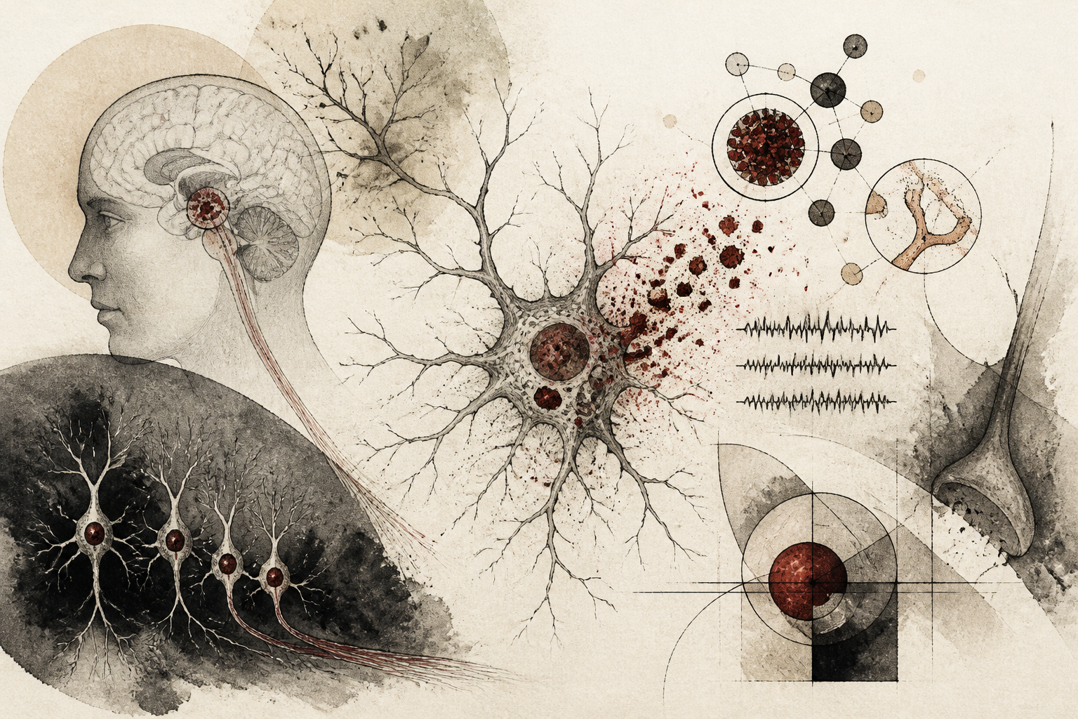
In the ongoing quest to decipher the molecular architecture of Parkinson’s disease, researchers have long grappled with the 'chicken or the egg' dilemma concerning neuroinflammation and neuronal death. While the loss of dopaminergic neurons in the substantia nigra pars compacta remains the clinical hallmark of the disease, the role of the brain’s resident immune cells, known as microglia, has frequently been viewed as a secondary response to injury. However, a groundbreaking study published in *Nature Neuroscience* suggests a more proactive, pathological role for these cells. The research identifies a deficiency in the protein NUS1 within microglia as a primary driver of dopaminergic neurodegeneration, mediated through a complex process of intercellular iron dysregulation. This discovery shifts the spotlight toward how the metabolic failures of immune cells can directly sabotage the survival of neighboring neurons, offering a new vantage point for therapeutic intervention.
The Genetic and Cellular Landscape of NUS1
To understand the implications of this study, one must first consider the physiological role of the NUS1 gene. NUS1 is known to encode a subunit of the cis-prenyltransferase complex, which is essential for protein glycosylation and the synthesis of dolichol. Prior genetic studies had already linked NUS1 mutations to various neurodegenerative conditions, including Parkinson’s disease and epilepsy. However, most research had focused on its function within neurons themselves. The novelty of the current investigation lies in its specific focus on microglia. As the primary defenders of the central nervous system, microglia are responsible for clearing debris, pruning synapses, and maintaining the chemical balance of the brain’s interstitial environment. When these cells undergo a loss of NUS1, the study reveals that their protective functions are not merely diminished; they become actively toxic. By utilizing sophisticated mouse models and human cellular data, the researchers demonstrated that microglial NUS1 deficiency initiates a cascade that eventually results in the selective death of the very neurons responsible for motor control.
Mechanisms of Intercellular Iron Dysregulation
The central finding of the research involves the mismanagement of iron, a vital but potentially volatile mineral. While iron is necessary for numerous enzymatic reactions and energy production in the brain, its levels must be strictly regulated to prevent oxidative stress. The researchers observed that in the absence of NUS1, microglia exhibit a significant disruption in their internal iron handling. Specifically, the deficiency leads to an abnormal accumulation of iron within the microglia, which in turn triggers a proinflammatory state. More critically, the study identified a breakdown in the communication between microglia and dopaminergic neurons regarding iron homeostasis. The dysfunctional microglia appear to promote an environment of 'iron overload' in the surrounding neural tissue. This excess iron facilitates the formation of reactive oxygen species through the Fenton reaction, leading to ferroptosis—a form of regulated cell death characterized by iron-dependent lipid peroxidation. The specificity of this effect on dopaminergic neurons is particularly noteworthy, as these cells are known to be naturally high in iron content and are thus uniquely vulnerable to any further metabolic shifts in their microenvironment.
Methodological Approaches and Findings
The research team employed a multi-faceted experimental design to isolate the effects of NUS1. They generated conditional knockout mice where the NUS1 gene was specifically deleted only in the microglial cells. These animals exhibited progressive motor deficits reminiscent of human Parkinson’s disease symptoms, such as impaired coordination and reduced locomotor activity. Histological analysis confirmed a significant reduction in the number of dopaminergic neurons in the substantia nigra of these mice compared to control groups. To validate these findings in a human context, the scientists analyzed single-nucleus RNA sequencing data from the brains of deceased Parkinson’s patients. They found that NUS1 expression was indeed significantly lower in the microglia of these patients, particularly in those areas most affected by the disease. Furthermore, the team conducted in vitro experiments using co-culture systems of microglia and neurons, demonstrating that the neurodegeneration could be mitigated by using iron chelators or by restoring NUS1 function, thereby proving that the iron-dependent pathway was the primary driver of the observed damage.
Limitations and Future Directions
While the study provides a compelling link between microglial health and neuronal survival, it also raises several questions that warrant further exploration. One limitation is the inherent difference between mouse models and the human brain; while the researchers used human data to support their claims, the temporal progression of Parkinson’s disease in humans spans decades, a timeline that is difficult to replicate in laboratory settings. Additionally, the precise molecular bridge between NUS1 loss and the physical movement of iron across cell membranes requires more granular mapping. Scientists are now left to wonder whether NUS1 deficiency is an early-stage trigger or a late-stage accelerant of the disease. Furthermore, it remains to be seen if this mechanism is exclusive to Parkinson’s or if it plays a broader role in other forms of neurodegeneration where iron accumulation is observed, such as Alzheimer’s disease or multiple sclerosis. Establishing the specificity of the NUS1-iron axis will be crucial for developing targeted therapies that do not inadvertently interfere with the essential functions of microglia in other regions of the brain.
Towards a New Therapeutic Paradigm
The significance of this research lies in its potential to transform the treatment landscape for Parkinson’s disease. Current therapies primarily focus on managing symptoms by replacing lost dopamine, rather than halting the underlying death of neurons. By identifying microglial NUS1 and iron dysregulation as key pathological drivers, this study points toward a 'disease-modifying' strategy. Interventions that can stabilize iron metabolism within the brain’s immune system or restore the functional integrity of the cis-prenyltransferase complex could theoretically protect dopaminergic neurons from oxidative damage before they are lost. This shift from neuron-centric models to a more holistic, multicellular understanding of brain health emphasizes that the support structures of the brain—the glia—are just as important as the neurons they protect. As the global population ages and the prevalence of Parkinson’s continues to rise, these molecular insights offer a necessary path forward in the search for a cure, moving us closer to a time when we can prevent the motor decline associated with this devastating disorder at its very source.
Quick answers
- What is the role of the NUS1 gene in the brain?
- NUS1 encodes a protein essential for the cis-prenyltransferase complex, which manages protein glycosylation. In the context of this study, its absence in microglia leads to iron dysregulation and neurotoxicity.
- How does iron contribute to Parkinson's disease progression?
- Excess iron in the brain can lead to oxidative stress and a specific type of cell death called ferroptosis. This study shows that dysfunctional microglia with NUS1 deficiency cause iron to accumulate dangerously near dopaminergic neurons.
- Could these findings lead to new Parkinson's treatments?
- Yes, by targeting microglial iron handling or the NUS1 pathway, researchers hope to develop therapies that protect neurons from dying, rather than just treating symptoms.
Rewritten by Zeit editorial AI. Based on original reporting at Nature Neuroscience.