Hormonal Longevity and Cerebrovascular Health: Estrogen’s Role in Aging
New longitudinal MRI research indicates that prolonged estrogen exposure significantly reduces the burden of cerebral small vessel disease in postmenopausal women.
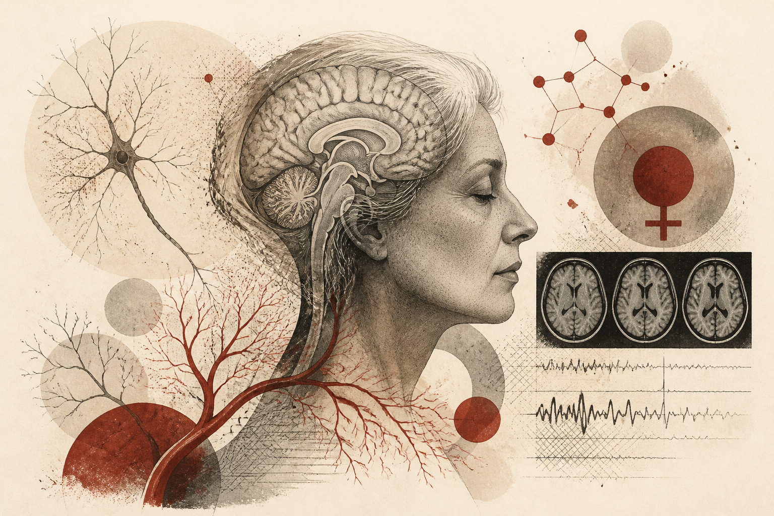
The biological mechanisms of neuroprotection have long prioritized the study of cardiovascular health and genetic predispositions, yet the specific impact of sex-specific hormonal history on the aging brain has remained an area of emerging inquiry. Recent longitudinal data published in the journal Neurology and reported by NeuroscienceNews suggests that the duration of a woman’s reproductive lifespan—and her history of exogenous hormone use—plays a critical role in preserving the structural integrity of the brain’s microcirculation. By analyzing the accumulation of white matter hyperintensities and other markers of cerebral small vessel disease, researchers have identified a significant correlation between high lifetime estrogen exposure and a lower risk of neurological decline in later life. This five-year study provides a robust framework for understanding how hormonal shifts over decades manifest as visible physiological changes in the aging cranium.
The Neurovascular Landscape of Small Vessel Disease
Cerebral small vessel disease (cSVD) is a major contributor to cognitive impairment, stroke, and physical frailty in older adults. It is primarily characterized by damage to the small arteries, arterioles, capillaries, and venules within the brain, often manifesting on magnetic resonance imaging (MRI) as white matter hyperintensities, lacunes, or microbleeds. For decades, the medical community has observed a sex-based discrepancy in the prevalence and severity of these markers as patients age. While men often exhibit higher rates of vascular complications in midlife, women frequently experience a sharp escalation in cerebrovascular burden following the menopausal transition. This shift has led scientists to hypothesize that the precipitous drop in estrogen—a steroid hormone with known vasodilatory and anti-inflammatory properties—may leave the female brain vulnerable to small vessel degradation.
The study featured in Neurology sought to quantify this protective effect by examining the cumulative exposure to estrogen over a woman's lifetime. Rather than viewing menopause as a singular event, the researchers looked at the 'reproductive window'—the years between menarche and menopause—as well as the supplemental influence of hormone therapy. The findings suggest that the brain's microcirculation is not merely a static system susceptible to wear and tear, but a dynamic network that may be actively shielded by the presence of circulating estrogens. This shielding effect appears to have long-term implications for white matter integrity, which is essential for the efficient transmission of signals between different regions of the brain.
Quantifying the Protective Window
To assess the relationship between hormones and brain health, the research team utilized high-resolution MRI scans over a five-year period, allowing them to track the progression of cSVD in real-time. The results revealed a striking disparity based on hormonal history. Participants with a longer reproductive lifespan—defined as more years spent in a premenopausal state—exhibited significantly lower volumes of white matter hyperintensities compared to those with shorter reproductive windows. Specifically, the data indicated that individuals with the highest levels of lifetime estrogen exposure saw a reduction in the burden of cerebral small vessel disease by as much as 59%. This suggests that every year of endogenous estrogen production contributes a cumulative 'deposit' into a woman's neurological reserve.
Furthermore, the study investigated the role of exogenous hormones, such as those used in hormone replacement therapy (HRT). The analysis showed that the benefits were not limited to natural reproductive longevity; the use of hormone therapy was also associated with a preserved microvascular profile. This aspect of the findings is particularly relevant given the historical controversy surrounding hormone therapy and vascular risk. By focusing specifically on the microcirculation of the brain over a five-year longitudinal span, this research clarifies that, for many, the hormonal intervention may serve as a critical buffer against the structural decay typically associated with aging and the postmenopausal state.
Mechanisms of Estrogen-Mediated Neuroprotection
The biological explanation for these findings lies in the multifaceted role estrogen plays within the vascular system. Estrogen receptors are widely distributed throughout the endothelial cells that line the blood vessels in the brain. When estrogen binds to these receptors, it promotes the production of nitric oxide, a molecule that facilitates vasodilation and helps maintain the elasticity of vessel walls. This ensures consistent blood flow and reduces the mechanical stress on small vessels. Additionally, estrogen exhibits potent anti-oxidative and anti-inflammatory effects, which prevent the cellular damage that leads to the scarring and leakage characteristic of white matter hyperintensities.
Methodologically, the five-year duration of the study allowed researchers to move beyond cross-sectional snapshots, which can often be misleading. By measuring the rate of change in brain lesions, they could confirm that estrogen does not just correlate with a 'healthier' brain at one point in time, but actually slows the rate of vascular accumulation as the individual ages. This distinction is vital for clinical psychology and neurology, as it suggests that hormonal health in the 40s and 50s creates a trajectory for cognitive health in the 70s and 80s. The preservation of white matter integrity is the direct result of a healthier, more resilient microvascular environment fostered by decades of hormonal activity.
Interpretations and Clinical Limitations
While the 59% reduction in cSVD burden is a significant figure, the interpretation of this data requires a nuanced understanding of individual health profiles. The researchers noted that while estrogen is a powerful variable, it does not exist in a vacuum. Other factors, such as blood pressure management, metabolic health, and genetic markers like the APOE4 allele, also influence the progression of small vessel disease. The study’s reliance on a specific cohort means that while the trends are clear, the exact magnitude of protection may vary across different populations. Moreover, the timing of hormone therapy—often referred to as the 'timing hypothesis'—suggests that the benefits may be most pronounced when therapy is initiated closer to the onset of menopause rather than in later senescence.
An open question remains regarding the threshold of estrogen exposure required to achieve these neuroprotective effects. Is there a point of diminishing returns, or does the protection scale linearly with every additional year of exposure? Furthermore, the study primarily focused on white matter hyperintensities. Future research will need to determine if this hormonal shield also prevents other forms of cSVD, such as microbleeds or perivascular space enlargement, and whether these structural benefits translate directly into a measurable reduction in clinical dementia cases. Understanding these limitations is essential for translating this academic finding into personalized medical advice.
Implications for Long-term Brain Health
Why does this research matter for the broader field of psychology and aging? It shifts the conversation from a reactive model of treating cognitive decline to a proactive model of vascular preservation. By identifying lifetime estrogen exposure as a key determinant of brain aging, healthcare providers can better identify at-risk populations and consider hormonal history as a critical component of a patient’s neurological profile. For women, this research validates the importance of reproductive health as a pillar of long-term cognitive stability.
As the global population ages and the prevalence of vascular-related cognitive impairment rises, the ability to protect the brain’s 'plumbing' becomes a public health priority. This study, sourced from the recent Neurology publication via NeuroscienceNews, reinforces the idea that the biological transitions of midlife have profound echoes in the neurological health of the elderly. By shielding the brain’s microcirculation, estrogen serves as a silent architect of the aging mind, maintaining the structural pathways necessary for cognitive function well into the later stages of life.
Quick answers
- How does estrogen protect the aging brain?
- Estrogen protects the brain by maintaining the health of small blood vessels, reducing white matter damage, and promoting better circulation through its anti-inflammatory and vasodilatory properties.
- What is the relationship between menopause and brain health?
- A shorter reproductive lifespan (earlier menopause) is linked to a higher burden of cerebral small vessel disease, whereas more years of estrogen exposure help preserve white matter integrity.
- Can hormone therapy help prevent brain aging?
- According to the study in Neurology, hormone therapy use was associated with lower levels of cerebral small vessel disease markers, suggesting it can supplement natural estrogen in protecting the brain's microcirculation.
Rewritten by Zeit editorial AI. Based on original reporting at NeuroscienceNews.com.