Early Volumetric Markers: Redefining the Timeline of Alzheimer's Degeneration
Recent findings from Johns Hopkins suggest that the amygdala and entorhinal cortex undergo measurable shrinkage years before traditional Alzheimer’s symptoms appear.
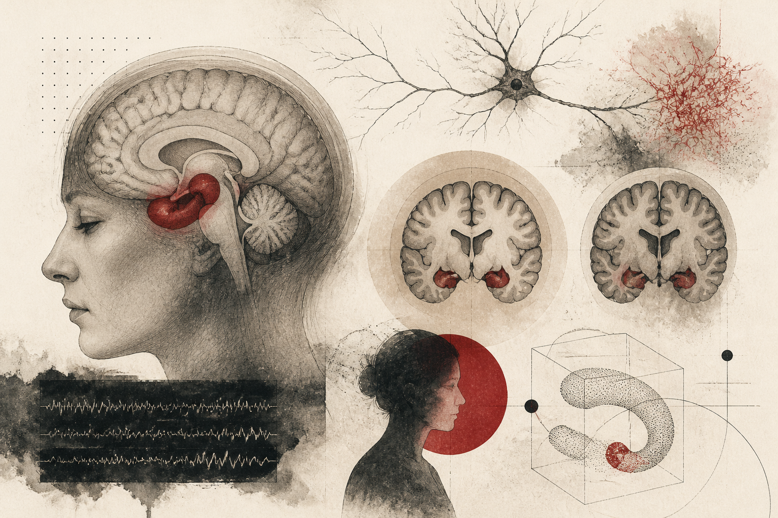
For decades, the clinical and diagnostic focus on Alzheimer’s disease has prioritized the hippocampus as the primary canary in the coal mine for neurodegeneration. This curved structure, essential for the consolidation of information from short-term to long-term memory, has long served as the gold standard for neuroimaging assessments. However, new research emerging from Johns Hopkins University suggests that the traditional diagnostic gaze may be shifting too late in the disease’s progression. According to a report originally published by ScienceDaily, researchers have identified subtle, hidden warning signs that manifest in the brain’s architecture up to a full decade before a patient experiences noticeable memory impairment. By shifting the focus away from the hippocampus toward the amygdala and the entorhinal cortex, scientists are uncovering a topographical map of decline that begins far earlier than previously understood.
The Topography of Early Neurodegeneration
The study conducted by the Johns Hopkins team utilized advanced neuroimaging to track the physical changes in the brains of participants over an extended period. Their findings indicate that the atrophy characteristic of Alzheimer’s disease does not begin simultaneously across all memory-related structures. Instead, the research highlights a specific sequence of decline where the amygdala—a region primarily associated with emotional processing—and the entorhinal cortex—a hub that serves as a gateway between the hippocampus and the neocortex—begin to shrink significantly earlier than the hippocampus itself. This revelation is significant because the hippocampus is often the first region clinicians inspect when a patient presents with cognitive complaints. By the time hippocampal shrinkage is visible on a standard MRI, the underlying pathological process may have already been underway for years, if not a decade.
This temporal gap in detection represents a critical window of opportunity. The Johns Hopkins scientists observed that the volumetric loss in the amygdala and entorhinal cortex preceded the onset of clinical memory loss, suggesting that the brain's emotional and sensory-integration centers are more vulnerable to the early stages of the disease than the central memory-forming apparatus. This chronological roadmap provides a new lens through which researchers can view the transition from healthy aging to preclinical Alzheimer’s, potentially moving the diagnostic threshold forward by several years.
Tau Protein and the Mechanism of Decay
To understand why these specific regions are targeted first, the researchers looked beyond simple volume measurements and investigated the biochemical drivers of the observed shrinkage. The study linked the early reduction in brain volume to the abnormal buildup of tau protein. While amyloid-beta plaques have historically dominated the discourse on Alzheimer’s pathology, tau tangles are increasingly recognized as the more accurate predictors of actual neuronal death and cognitive decline. The Johns Hopkins research confirms that the presence of these toxic tau proteins correlates directly with the structural thinning of the amygdala and entorhinal cortex.
Tau protein, in its healthy state, stabilizes the internal microtubules of neurons, ensuring the smooth transport of nutrients and signals. In Alzheimer’s, however, these proteins collapse into twisted strands called tangles, which disrupt the cell's transport system and eventually lead to cell death. The specific vulnerability of the entorhinal cortex to early tau accumulation has been noted in previous neuropathological studies, but the Johns Hopkins team’s ability to correlate this with measurable volumetric shrinkage in living patients—years before memory loss—bridges the gap between molecular biology and clinical observation. This mechanism suggests that the shrinkage is not a random byproduct of aging but a direct consequence of protein-mediated neurotoxicity targeting specific neural circuits.
Redefining Diagnostic Boundaries and Limitations
While these findings offer a promising new pathway for early detection, they also highlight the limitations of current diagnostic paradigms. The traditional reliance on hippocampal volume may be resulting in a high rate of 'late' diagnoses, where interventions are applied only after significant neural damage has occurred. By recognizing the amygdala and entorhinal cortex as earlier indicators, the medical community may be able to identify high-risk individuals while their cognitive functions are still intact. However, the researchers caution that brain shrinkage is a complex phenomenon. While the correlation between these specific regions and tau buildup is strong, other factors such as chronic stress, cardiovascular health, and environmental influences can also impact brain volume.
Furthermore, the study raises questions regarding the specificity of these markers. Does shrinkage in the amygdala always signal Alzheimer’s, or could it indicate other neurodegenerative or psychiatric conditions? The Johns Hopkins team emphasizes that these volumetric changes should be viewed as part of a larger diagnostic picture, integrated with genetic testing and other biomarkers. Another limitation involves the accessibility of the technology required to measure these subtle changes; high-resolution imaging and sophisticated volumetric analysis software are not yet standard in every clinical setting, which may delay the widespread adoption of these early-detection methods.
The Clinical Imperative for Early Intervention
The implications of this research extend far beyond the laboratory. If the onset of Alzheimer’s can be predicted a decade in advance, the window for lifestyle interventions and experimental therapies expands dramatically. Current pharmacological treatments for Alzheimer’s often struggle to show efficacy in late-stage patients because the sheer volume of lost neurons is too great to overcome. An earlier diagnosis, facilitated by monitoring the amygdala and entorhinal cortex, could allow for the deployment of tau-targeting drugs at a point where they can preserve existing brain function rather than attempting to restore what is already gone.
Moreover, this research underscores the importance of the emotional and sensory components of brain health. The early involvement of the amygdala suggests that subtle changes in emotional regulation or mood might be early behavioral markers of the disease, occurring in tandem with the physical shrinkage. As the scientific community continues to parse these findings, the goal remains clear: to move away from a reactive model of Alzheimer’s care toward a proactive, preventative strategy. By listening to the early warning signs emitted by the brain's emotional and integrative centers, clinicians may finally gain the upper hand in the fight against this progressive cognitive decline. The Johns Hopkins study provides the necessary evidence that the journey toward Alzheimer’s starts long before the first memory fades, and identifying those first steps is the key to eventually halting the march of the disease.
Quick answers
- Which brain regions shrink first in Alzheimer's disease according to new research?
- Research from Johns Hopkins indicates the amygdala and entorhinal cortex shrink up to a decade before the hippocampus and the onset of memory loss.
- How long before memory loss can Alzheimer's signs be detected?
- Hidden signs such as volumetric shrinkage in specific brain regions can be detected up to 10 years before clinical memory symptoms appear.
- What causes the brain shrinkage associated with early Alzheimer's?
- The shrinkage is linked to the abnormal buildup of tau protein, which forms tangles that lead to neuronal death and structural thinning.
Editorial Team