Early Cortical Thinning as a Precursor to Amyloid-Beta Accumulation in Alzheimer’s Disease
New longitudinal research published in Nature Neuroscience suggests that structural changes in the brain’s cortex may precede significant amyloid buildup by nearly a decade.
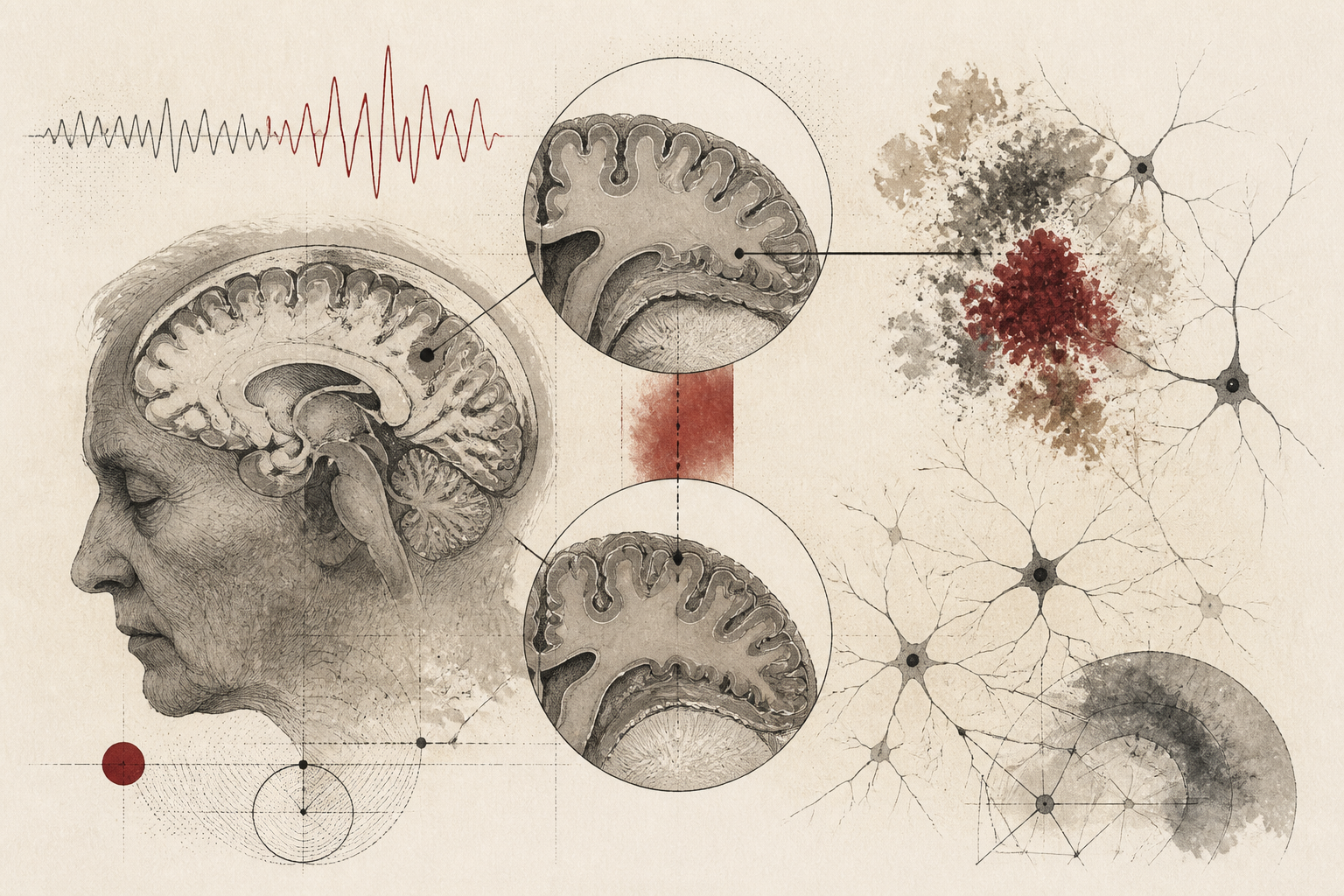
The trajectory of Alzheimer’s disease research has long been anchored by the amyloid cascade hypothesis, which posits that the accumulation of amyloid-beta plaques is the primary driver of subsequent neurodegeneration. However, a significant paradigm shift may be underway following a comprehensive longitudinal study published in Nature Neuroscience. Researchers have identified that specific changes in cortical thickness do not merely coincide with the presence of amyloid-beta; rather, these structural alterations appear to precede high levels of protein accumulation by at least seven years. This discovery suggests that the earliest phases of the disease might be detectable through neuroimaging far earlier than previously understood, potentially redefining the window for therapeutic intervention.
Traditionally, clinicians have relied on the detection of amyloid-beta via positron emission tomography (PET) scans or cerebrospinal fluid analysis as the definitive hallmark of the preclinical stage of Alzheimer's. While these biomarkers remain critical for diagnosis, the temporal relationship between protein deposition and actual brain atrophy has remained a subject of intense academic debate. The recent findings suggest that the brain undergoes subtle but measurable structural changes long before the threshold for amyloid positivity is met. By tracking these minute variations in the cortical mantle, the research team has provided a new timeline for the biological progression of cognitive decline, emphasizing a protracted period of vulnerability that precedes traditional diagnostic markers.
Methodological Framework and Longitudinal Tracking
The study’s findings are the result of rigorous longitudinal tracking involving a cohort of individuals who underwent repeated neuroimaging over several years. By utilizing advanced magnetic resonance imaging (MRI) techniques, the researchers were able to measure the thickness of various cortical regions with high precision. These measurements were then cross-referenced with PET scan data to monitor the accumulation of amyloid-beta plaques over the same period. The methodology allowed the team to map the rate of cortical thinning against the rate of protein deposition, creating a high-resolution temporal map of the aging brain.
One of the most significant aspects of the study’s design was its focus on the 'pre-amyloid' phase. By enrolling participants who were initially below the clinical threshold for amyloid positivity, the researchers could observe the transition from a healthy state to the earliest signs of pathology. This approach differs from cross-sectional studies, which often only capture a snapshot of the disease at a single point in time. The longitudinal nature of the data ensured that each participant served as their own control, allowing the team to filter out individual differences in brain structure and focus purely on the rate of change over time. This granular level of detail was essential in establishing the seven-year lead time between cortical thinning and the surge in amyloid levels.
The Sequence of Neurobiological Degradation
Central to the study is the observation that cortical thickness in specific regions—most notably those associated with memory and executive function—begins to decline well before amyloid-beta reaches levels commonly associated with Alzheimer’s pathology. The data indicates that this thinning is not a localized event but follows a systematic pattern across the brain’s architecture. As the cortex loses density, there appears to be a dormant period of approximately seven years before the rapid accumulation of amyloid plaques is detected. This sequence challenges the assumption that amyloid-beta is the sole initiator of the degenerative process.
The mechanisms behind this early thinning remain a subject of investigation, but the research points toward a complex interplay of factors. It is possible that early-stage cellular dysfunction or metabolic changes are responsible for the initial loss of cortical volume, which then creates a biological environment conducive to the deposition of amyloid-beta. Alternatively, the thinning could represent a separate, parallel process of neurodegeneration that simply manifests earlier than the visible buildup of protein aggregates. Regardless of the underlying trigger, the consistency of this seven-year gap across the study’s cohort suggests a highly predictable biological cadence in the progression of the disease.
Implications for Clinical Interpretation and Diagnostics
The interpretation of these findings has profound implications for the future of geriatric medicine and neurology. If cortical thinning is indeed a reliable precursor to amyloid accumulation, MRI could become a more prominent tool in the early screening process. Currently, PET scans and lumbar punctures are the primary methods for detecting amyloid, but these can be expensive or invasive. In contrast, MRI is widely available and non-invasive, making it a viable candidate for large-scale population screening. By identifying individuals who exhibit accelerated cortical thinning, clinicians might be able to flag at-risk patients nearly a decade before they would typically test positive for amyloid-beta.
Furthermore, this research shifts the focus toward the importance of 'baseline' neuroimaging. Understanding an individual’s normal rate of cortical change is necessary to distinguish between healthy aging and the early stages of neurodegeneration. The study highlights that the absolute thickness of the cortex is often less important than the trajectory of change. A patient may still have a 'normal' cortical thickness relative to the general population but could be experiencing an abnormal rate of decline that signals future pathology. This nuanced understanding of brain morphology requires a move away from static diagnostic thresholds toward a more dynamic, longitudinal model of patient care.
Limitations and Future Research Directions
Despite the groundbreaking nature of the study, the researchers acknowledge several limitations that must be addressed in subsequent investigations. First, while the seven-year window was observed in a significant portion of the cohort, there is still variability in how the disease manifests across different individuals. Factors such as genetic predisposition, lifestyle, and cardiovascular health may influence the speed at which cortical thinning leads to amyloid buildup. Therefore, the findings should be viewed as a general biological trend rather than a universal rule for every patient.
Another open question involves the role of tau proteins, which are also characteristic of Alzheimer’s disease. This specific study focused primarily on the relationship between cortical thickness and amyloid-beta, but the interplay between structural atrophy and tau pathology is another critical piece of the puzzle. Future studies will need to incorporate tau imaging to determine if the early cortical thinning is also linked to the spread of neurofibrillary tangles. Additionally, the researchers call for more diverse cohorts to ensure that these temporal patterns hold true across different ethnicities and socioeconomic backgrounds, as most current data is derived from relatively homogeneous populations.
The Future of Preventative Intervention
Why does this research matter for the average person and the scientific community at large? The answer lies in the 'therapeutic window.' Many clinical trials for Alzheimer’s drugs have failed because the interventions were administered too late in the disease process, after significant and irreversible brain damage had already occurred. By identifying a marker that appears seven years before amyloid-beta peaks, scientists have opened a new door for preventative medicine. If a drug or lifestyle intervention could be introduced at the first sign of cortical thinning, it might be possible to delay or even prevent the subsequent accumulation of amyloid-beta.
This study, published in Nature Neuroscience, serves as a reminder that the brain is a highly dynamic organ and that the transition to disease is a slow, multi-decade process. By refining our ability to see the earliest ripples of change, we move closer to a future where Alzheimer’s is not a diagnosis of late-stage decline, but a manageable condition detected and treated in its infancy. The goal is no longer just to treat the symptoms of the disease, but to understand and interrupt the biological countdown long before the first symptoms ever appear.
Quick answers
- What is the main finding of the Nature Neuroscience study regarding Alzheimer's?
- The study found that measurable thinning of the brain's cortex occurs at least seven years before amyloid-beta protein levels reach the high thresholds typically associated with Alzheimer's disease.
- Why is cortical thinning important for Alzheimer's diagnosis?
- It suggests that structural brain changes detected by MRI could serve as a much earlier warning sign for neurodegeneration than current amyloid-based biomarkers, potentially offering a longer window for intervention.
- Does this study mean amyloid is not the cause of Alzheimer's?
- Not necessarily. It indicates that the sequence of the disease is more complex than previously thought, with structural atrophy preceding the measurable buildup of amyloid plaques, suggesting they are parts of a longer, interconnected process.
Rewritten by Zeit editorial AI. Based on original reporting at Nature Neuroscience.