Bone-Brain Signaling: How Tibial Compression Facilitates Post-Injury Neurological Recovery
Recent findings in Nature Neuroscience reveal that mechanical stimulation of long bones activates a PIEZO1-mediated pathway, accelerating recovery following traumatic brain injury.
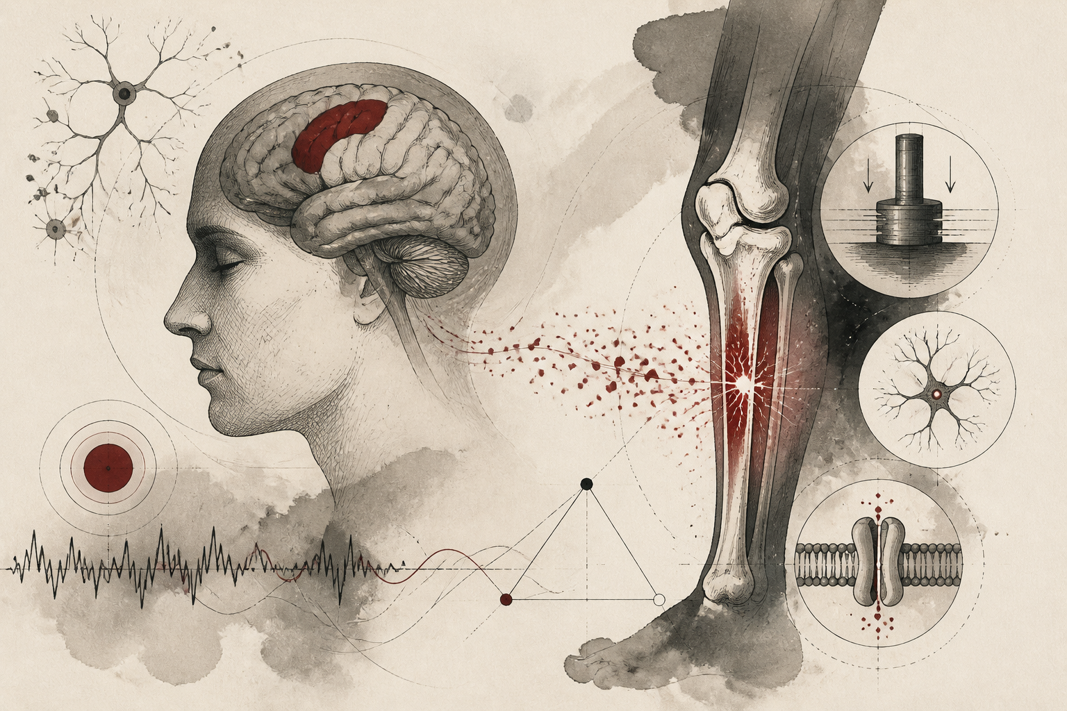
The relationship between physical activity and cognitive health has long been established in clinical literature, yet the specific biological mechanisms connecting mechanical bodily stress to neural repair have remained elusive. For decades, researchers have observed that patients who engage in early, controlled physical mobilization following traumatic brain injury (TBI) often exhibit superior recovery outcomes compared to those remains sedentary. While general cardiovascular health was once thought to be the primary driver of this phenomenon, a groundbreaking study published in *Nature Neuroscience* titled "Tibial bone compression promotes recovery after brain injury through osteocyte PIEZO1" suggests a more direct, endocrine-like communication between the skeletal system and the central nervous system. This research shifts the focus from the lungs and heart to the bones, identifying a specific mechanosensitive pathway that translates physical pressure on the legs into restorative signals for the brain.
The Skeletal System as an Endocrine Organ
Traditional anatomy often categorizes the skeletal system as a passive framework designed for structural support and mineral storage. However, emerging research over the last two decades has redefined bone as an active endocrine organ capable of secreting hormones, such as osteocalcin, which influence metabolism and brain function. The study in *Nature Neuroscience* builds upon this paradigm by examining how mechanical loading—specifically the compression of the tibia, or shinbone—impacts the brain's internal environment following a traumatic insult. The researchers focused on osteocytes, the most abundant cell type in mature bone tissue, which act as primary sensors for mechanical strain. By utilizing sophisticated murine models of brain injury, the investigative team sought to determine if the physical act of bone compression could trigger a systemic response robust enough to cross the blood-brain barrier and facilitate tissue repair.
Mechanisms of Mechanosensation: The Role of PIEZO1
At the heart of this discovery is PIEZO1, a specialized mechanosensitive ion channel that converts physical pressure into biological signals. The researchers identified that when the tibia undergoes compression, PIEZO1 channels within osteocytes are activated. This activation initiates a cascade of intracellular events that results in the secretion of specific signaling molecules into the bloodstream. The study demonstrated that in models where PIEZO1 was genetically deleted from osteocytes, the neuroprotective effects of bone compression were entirely abolished. This suggests that the bone-to-brain recovery axis is not a generalized byproduct of exercise, but a precise molecular response triggered by the mechanical deformation of bone tissue itself. Once these bone-derived signals reach the brain, they appear to modulate the activity of microglia—the brain's resident immune cells—shifting them from a pro-inflammatory state to a pro-repair state, thereby reducing secondary damage following a TBI.
Experimental Findings and Neurological Outcomes
To validate these findings, the research team employed a series of controlled experiments where mice with brain injuries were subjected to daily sessions of non-invasive tibial compression. These sessions were designed to mimic the mechanical forces experienced during weight-bearing activities like walking or standing. The results were striking: the mice receiving bone stimulation showed significantly reduced lesion volumes and improved motor coordination compared to the control group. Furthermore, cognitive assessments revealed that the stimulated mice regained spatial memory functions at a faster rate. The researchers also noted an increase in the expression of neurotrophic factors, which are essential for the survival and growth of neurons. These findings provide a compelling biological explanation for why early mobilization in clinical settings is so effective, emphasizing that the simple act of putting weight on the limbs may be sending essential instructions to the recovering brain.
Interpretations and Clinical Implications
These findings suggest that the skeletal system acts as a critical mediator of neural plasticity. The discovery that tibial compression can influence brain recovery opens new avenues for rehabilitative medicine, particularly for patients who are unable to engage in vigorous aerobic exercise. If mechanical stimulation of the bone alone can trigger the PIEZO1 pathway, it may be possible to develop specialized medical devices that provide targeted bone compression for bedridden patients or those with severe motor impairments. This "bottom-up" approach to neuroprotection—targeting the limbs to save the brain—challenges the traditional "top-down" view of neurological treatment. It suggests that the body's internal systems are far more integrated than previously understood, with the skeleton acting as a sentinel that monitors physical activity levels to regulate the brain's capacity for self-repair.
Limitations and Future Research Directions
While the study provides a robust framework for understanding the bone-brain axis, several limitations must be addressed before these findings can be fully translated to human clinical practice. The primary research was conducted using murine models, and while the PIEZO1 channel is conserved across species, the human skeletal system's scale and complexity may produce different signaling magnitudes. Furthermore, the optimal "dose" of bone compression—frequency, intensity, and duration—remains to be determined. Questions also remain regarding the specific identities of all the bone-derived factors (osteokines) involved in this process. Future studies will need to investigate whether other bones, such as the femur or ulna, contribute similarly to this effect and whether this pathway can be leveraged to treat chronic neurodegenerative conditions like Alzheimer’s disease or stroke, where neural repair is also a primary goal.
Integrating Bone Health into Neurorehabilitation
The significance of this research lies in its potential to revolutionize post-injury care protocols. By identifying the osteocyte PIEZO1 pathway, the study provides a biological imperative for prioritizing skeletal health in neurological patients. It reinforces the idea that the brain does not heal in isolation; it requires input from the rest of the body to optimize its recovery processes. As Zeit Psychology Online University continues to monitor developments in neurobiology, this study stands as a testament to the interdisciplinary nature of modern science, bridging the gap between orthopedics and neuroscience. For clinicians, the message is clear: to heal the mind, one must not overlook the importance of the bones. The future of brain injury recovery may well depend on our ability to harness the hidden signals generated within our very foundations.
Quick answers
- How does bone compression affect the brain according to recent research?
- Mechanical compression of the tibia activates PIEZO1 ion channels in bone cells (osteocytes), which then release signals that travel to the brain to promote repair and reduce inflammation after injury.
- What is PIEZO1 and why is it important for recovery?
- PIEZO1 is a mechanosensitive ion channel that detects physical pressure. In the context of brain injury, it acts as a trigger in the bones to start a chemical signaling process that aids neurological healing.
- Can bone stimulation replace exercise for brain health?
- While the study shows bone compression alone has benefits, it is currently viewed as a potential supplement to exercise or an alternative for patients who cannot move, rather than a total replacement for the cardiovascular benefits of physical activity.
Rewritten by Zeit editorial AI. Based on original reporting at Nature Neuroscience.