Amygdala Neuron Specialization and Its Role in Human Episodic Memory Encoding
Researchers identify stimulus-specific neurons in the human amygdala that predict how successfully we form new episodic memories.
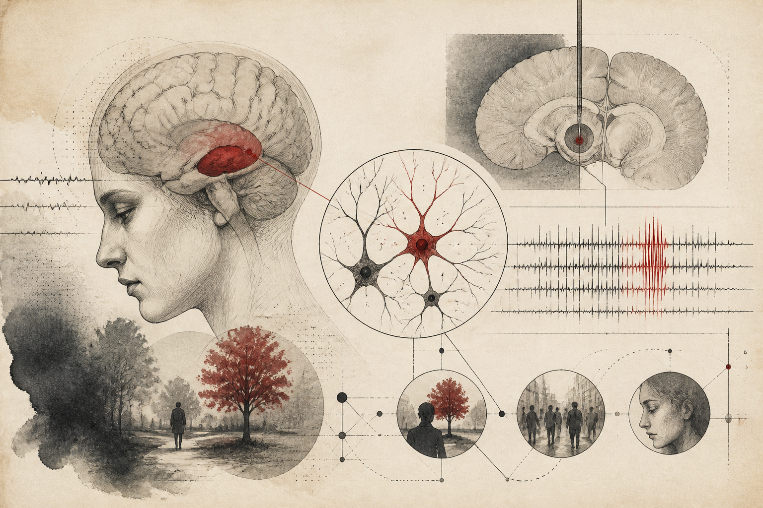
In the complex landscape of the human brain, the mechanisms by which transient experiences are transformed into lasting memories remain a focal point of neuroscientific inquiry. A recent study published in Nature Neuroscience has provided significant clarity on this process, identifying a specific population of neurons within the amygdala that appear to govern the success of episodic memory encoding. By observing the firing patterns of individual cells in human subjects, researchers have demonstrated that the amygdala is not merely a center for emotional processing, but a sophisticated regulator of mnemonic fidelity, particularly when processing specific visual stimuli.
Traditional models of memory often place the hippocampus at the center of the encoding process. However, the interplay between the medial temporal lobe structures and the amygdala has long suggested a more integrated system. This new research advances our understanding by demonstrating that the recruitment of stimulus-specific neurons in the amygdala can predict whether a person will successfully remember a specific image later. The findings suggest that the amygdala acts as a selective filter, highlighting relevant information to ensure it is prioritized by the brain’s memory-forming machinery.
Cellular Dynamics and Encoding Efficacy
The study’s findings center on the discovery of "stimulus-specific" neurons. Unlike generalized neurons that might fire in response to any visual input, these cells are highly selective, responding only to specific categories or features of a stimulus. The researchers found that when these specialized neurons in the amygdala showed high levels of activity during the initial exposure to an image, the likelihood of the subject remembering that image during subsequent testing increased significantly. This correlation suggests that the amygdala’s internal representation of a stimulus is a critical precursor to the stabilization of a memory trace.
Furthermore, the data indicates that this predictive power is not universal across all neuron types. The study distinguished between general arousal-related activity and the specific, tuned responses of individual cells. It was the precise recruitment of these tuned cells that served as the most reliable indicator of encoding success. This nuance is vital, as it shifts the academic conversation away from the amygdala as a blunt instrument of emotional intensity toward a role involving high-resolution information processing. The specificity of these neurons implies that the amygdala is deeply involved in identifying the 'what' of an experience, rather than just the 'how important' or 'how scary' aspects.
Methodological Precision in Human Electrophysiology
Investigating the firing patterns of individual neurons in living humans requires extraordinary methodological rigor. The researchers utilized intracranial recordings from patients undergoing clinical monitoring for epilepsy. This setup allowed the team to record single-neuron activity—a level of resolution that non-invasive techniques like fMRI cannot achieve. During the experiments, participants were shown a series of images while their neural activity was monitored. A subsequent memory test categorized these images as either 'remembered' or 'forgotten,' allowing the researchers to look back at the neural data from the initial encoding phase to find patterns distinguishing the two outcomes.
The use of single-unit recording is particularly significant because it allows for the differentiation of local circuit dynamics. By analyzing the timing and frequency of spikes in individual amygdala neurons, the researchers could map the relationship between neural firing and cognitive performance. This method provides a direct link between cellular physiology and behavioral outcomes, bypassing the proxy measures of blood flow or broad electromagnetic fields. The precision of this approach reinforces the validity of the findings, suggesting that the observed stimulus-specific recruitment is a fundamental property of the human memory system.
Implications for Neurological Models and Memory Disorders
The interpretation of these results has wide-reaching implications for our theoretical models of the medial temporal lobe. If the amygdala provides a stimulus-specific boost to encoding, it may function as a bridge between sensory perception and long-term storage. This suggests that the amygdala provides a qualitative tag to experiences, signaling to the hippocampus and cortex that a specific set of information warrants preservation. This 'tagging' mechanism explains why certain vivid details of an event are retained while the surrounding context may fade.
From a clinical perspective, understanding the amygdala's role in memory encoding offers potential insights into disorders where memory is compromised or pathologically hyper-active. For instance, in conditions like Post-Traumatic Stress Disorder (PTSD), the over-recruitment of these stimulus-specific neurons might explain the intrusive and overly vivid nature of traumatic memories. Conversely, in neurodegenerative diseases like Alzheimer’s, a failure in the recruitment of these neurons could contribute to the inability to form new episodic memories. By identifying the specific cellular drivers of encoding, researchers may eventually develop targeted interventions—pharmacological or electrical—to modulate memory strength.
Limitations and Future Directions
While the study provides a robust framework for understanding the amygdala's role in memory, several open questions remain. The most prominent limitation is the specific population studied: patients with epilepsy. While these patients provide a unique opportunity for intracranial recording, their underlying neurological conditions could potentially influence neural dynamics. However, the researchers took care to record from regions outside the primary seizure focus to mitigate this risk. Additionally, the study focused primarily on visual stimuli; whether the same stimulus-specific recruitment occurs for auditory, olfactory, or abstract conceptual information remains to be seen.
Another area for future investigation is the directionality of the signaling. While the study shows that amygdala activity predicts memory, the precise temporal relationship between the amygdala and the hippocampus during these moments of successful encoding needs further exploration. Does the amygdala lead the hippocampus, or is there a bidirectional dialogue that occurs in the milliseconds following stimulus exposure? Answering these questions will require even more sophisticated multi-region recordings and computational modeling to map the flow of information across the brain’s memory circuits.
Why It Matters
This research represents a significant step forward in human neuroscience by pinpointing the cellular basis of memory formation. By moving beyond the idea of the amygdala as a simple "fear center," the study highlights its role as a sophisticated processor of specific information. This shift in perspective is essential for developing a more accurate map of the human mind and its capacity for learning. In an era where cognitive enhancement and the treatment of memory-related pathologies are becoming increasingly relevant, identifying the specific neurons responsible for successful encoding provides a concrete target for future scientific and medical progress. The ability to predict memory success at the level of a single cell underscores the incredible precision of the human brain and the ongoing potential of neuroscience to unlock its deepest mysteries.
Quick answers
- What is the main finding of the Nature Neuroscience study on the amygdala?
- The study found that specific neurons in the human amygdala, which respond to particular visual stimuli, fire more intensely when a person successfully encodes a memory they will later remember.
- How does the amygdala contribute to memory beyond emotional processing?
- Research indicates the amygdala acts as a stimulus-specific filter that identifies important information and signals the brain to prioritize it for long-term storage, rather than just processing fear or arousal.
- Why was the use of single-neuron recording important in this memory study?
- It allowed researchers to see the activity of individual cells in real-time, proving that specific 'tuned' neurons—not just general brain activity—are responsible for predicting memory success.
Rewritten by Zeit editorial AI. Based on original reporting at Nature Neuroscience.