Acoustic Attenuation and Motion Fidelity in Murine Functional Magnetic Resonance Imaging
Researchers introduce SORDINO, a novel fMRI methodology that eliminates acoustic noise and motion artifacts in awake mouse models to enhance neuroscientific data quality.
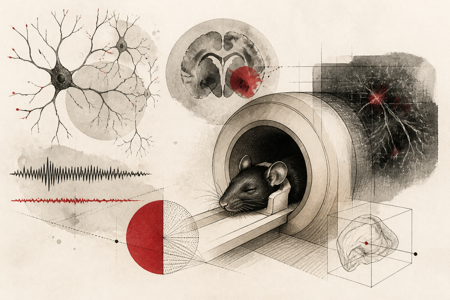
Traditional functional magnetic resonance imaging (fMRI) has long served as a cornerstone of cognitive neuroscience, yet its application in preclinical murine models has been historically constrained by the physical environment of the scanner itself. In a significant methodological advancement recently detailed in Nature Neuroscience, researchers have introduced SORDINO (silent, sensitive, specific, and artifact-resistant fMRI in awake, behaving mice). This novel approach addresses the dual challenges of acoustic distress and motion-induced signal distortion, offering a clearer window into the neural correlates of behavior without the confounding influence of pharmacological sedation or environmental noise. By reconciling the technical limitations of high-field imaging with the physiological requirements of awake subjects, SORDINO represents a pivot point for longitudinal studies in behavioral neuroscience.
The Challenge of the Awake Preclinical Model
The necessity of studying the brain in an awake state is driven by the fact that anesthesia fundamentally alters neurovascular coupling and global connectivity patterns. While early fMRI research relied heavily on sedated subjects to minimize movement, these pharmacological agents suppress the very cognitive processes researchers seek to observe. However, transitioning to awake subjects introduces a significant methodological paradox: the extreme acoustic noise generated by the rapid switching of magnetic gradients—often exceeding 100 decibels—induces a chronic stress response in rodents. This stress response alters heart rate, corticosterone levels, and baseline neural activity, potentially invalidating observations regarding natural behavior. Furthermore, even minute head movements or jaw motions in awake mice produce significant susceptibility artifacts that can be mistaken for neural activation or result in the total loss of usable data points.
Methodological Innovation and Acoustic Control
The SORDINO framework, as reported in Nature Neuroscience, utilizes a specialized pulse sequence and hardware configuration designed to achieve near-silent operation. Unlike standard echo-planar imaging (EPI) sequences that produce the characteristic loud rhythmic thumping, SORDINO employs a silent imaging strategy that reduces the acoustic pressure to levels indistinguishable from the ambient laboratory background. This is achieved through the optimization of gradient trajectories that avoid the resonance frequencies of the scanner bore. The reduction in noise not only improves animal welfare but also ensures that the sensory processing observed during experiments is not contaminated by the brain's response to the scanner’s internal environment. By stabilizing the baseline physiological state of the mouse, the researchers have created a controlled environment where specific stimuli can be presented and analyzed with unprecedented clarity.
Mechanisms of Artifact Suppression and Sensitivity
Beyond its acoustic benefits, SORDINO addresses the perennial issue of motion artifacts through a combination of hardware stabilization and advanced signal processing. The research team implemented a refined head-fixation system that integrates seamlessly with the RF coil, minimizing the displacement of the subject. Crucially, the SORDINO sequence is inherently resistant to the magnetic field inhomogeneities typically caused by movement. In traditional fMRI, even the act of breathing or minor muscle twitches can distort the magnetic field, leading to spatial blurring or signal dropouts. The SORDINO method utilizes a radial sampling technique in k-space, which is naturally more robust against such perturbations than the Cartesian sampling used in standard EPI. This results in a higher signal-to-noise ratio (SNR) and allows for the detection of subtle hemodynamic changes in small subcortical structures that were previously obscured by noise.
Interpretation of Enhanced Specificity
The specificity of the SORDINO method was validated through a series of behavioral tasks where mice were required to respond to sensory cues. The data indicated that the neural activations mapped via SORDINO were more tightly correlated with the known anatomical boundaries of functional regions compared to traditional methods. This enhancement in spatial specificity allows researchers to distinguish between adjacent cortical columns and tiny thalamic nuclei. The researchers noted that the hemodynamic response functions captured by SORDINO align more closely with simultaneous electrophysiological recordings, suggesting that the method provides a more faithful representation of underlying neuronal firing. This alignment is critical for translational research, as it bridges the gap between cellular-scale observations and macro-scale brain mapping.
Limitations and Future Directions
Despite the clear advantages of the SORDINO technique, the researchers acknowledge several limitations that warrant further investigation. The temporal resolution of the current sequence, while sufficient for most hemodynamic studies, still lags behind the millisecond precision of direct electrical recordings. Additionally, the implementation of SORDINO requires specialized gradient hardware and software modifications that may not be immediately available in all preclinical imaging centers. There is also the question of how this method scales with different magnetic field strengths; while the study demonstrated success at high fields, its performance at lower field strengths remains to be fully characterized. Future research will likely focus on integrating SORDINO with optogenetic tools to allow for the simultaneous stimulation and imaging of specific neural circuits without the interference of metal-based electrodes.
Implications for Neuropsychiatric Research
The development of SORDINO holds profound implications for the study of neuropsychiatric disorders. Conditions such as anxiety, post-traumatic stress disorder, and sensory processing disorders are difficult to model in anesthetized animals or in environments characterized by extreme noise. By providing a silent and stable imaging environment, SORDINO enables the longitudinal study of disease progression in mouse models that more accurately mirror the human experience. As the field moves toward more personalized and precise models of brain function, the ability to observe the awake, behaving brain without the interference of the scanning process itself will be an essential tool for the next generation of neuroscientists. This methodological leap, attributed to the work published in Nature Neuroscience, ensures that the data collected in preclinical trials is as robust and translatable as possible.
Quick answers
- What is SORDINO in the context of brain imaging?
- SORDINO is a specialized fMRI methodology designed for awake mice that eliminates scanner noise and reduces motion artifacts, allowing for more accurate neural data.
- Why is it important to image mice while they are awake?
- Anesthesia suppresses normal brain activity and changes how blood flows in response to neural triggers, so awake imaging is necessary to study natural behavior and cognition.
- How does SORDINO handle the loud noise of an MRI machine?
- It uses optimized magnetic gradient trajectories that avoid resonance, resulting in a silent scanning process that reduces stress in the animal subjects.
Rewritten by Zeit editorial AI. Based on original reporting at Nature Neuroscience.